Foramen spinosum

Last updated
Foramen spinosum
Gray145.png
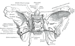
The inner surface of the sphenoid bone, with the foramen spinosum labelled at the left, second from the bottom.
Gray193.png
The inner surface of the base of the skull, with the sphenoid bone yellowed. The foramen spinosum is visible at the bottom right of the bone.
Details
Part of sphenoid bone
System skeletal
Identifiers
Latin foramen spinosum
TA98 A02.1.05.038
TA2 624
FMA 53156
Anatomical terms of bone

The foramen spinosum is a small open hole in the greater wing of the sphenoid bone that gives passage to the middle meningeal artery and vein, and the meningeal branch of the mandibular nerve (sometimes it passes through the foramen ovale instead).

Contents

The foramen spinosum is often used as a landmark in neurosurgery due to its close relations with other cranial foramina. It was first described by Jakob Benignus Winslow in the 18th century.

Structure

The foramen spinosum is a small foramen in the greater wing of the sphenoid bone of the skull. [1] :509 It connects the middle cranial fossa (superiorly), and infratemporal fossa (inferiorly). [2]

Contents

The foramen transmits the middle meningeal artery and vein, [1] :442 and sometimes the meningeal branch of the mandibular nerve (it may pass through the foramen ovale instead). [1] :364, 496

Relations

Inner surface of the base of skull, showing cranial foramina Skull foramina labeled.svg
Inner surface of the base of skull, showing cranial foramina

The foramen is situated just anterior to the sphenopetrosal suture. [1] :509 It is located posterolateral to the foramen ovale, and anterior to the sphenoidal spine. [2]

A groove for the middle meningeal artery and vein extends anterolaterally from the foramen. [1] :509

Variation

The foramen spinosum varies in size and location. The foramen is rarely absent, usually unilaterally, in which case the middle meningeal artery enters the cranial cavity through the foramen ovale. [3] It may be incomplete, which may occur in almost half of the population. Conversely, in a minority of cases (less than 1%), it may also be duplicated, particularly when the middle meningeal artery is also duplicated. [3] [4]

The foramen may pass through the sphenoid bone at the apex of the spinous process, or along its medial surface. [4]

Development

In the newborn, the foramen spinosum is about 2.25 mm long and in adults about 2.56 mm. The width of the foramen variesfrom 1.05 mm to about 2.1 mm in adults. [5] The average diameter of the foramen spinosum is 2.63 mm in adults. [6] It is usually between 3 and 4 mm away from the foramen ovale in adults. [7]

The earliest perfect ring-shaped formation of the foramen spinosum was observed in the eighth month after birth and the latest seven years after birth in a developmental study of the foramen rotundum, foramen ovale and foramen spinosum. The majority of the foramina in the skull studies were round in shape. [6] The sphenomandibular ligament, derived from the first pharyngeal arch and usually attached to the spine of the sphenoid bone, may be found attached to the rim of the foramen. [4] [8]

Animals

In other great apes, the foramen spinosum is found not in the sphenoid, but in parts of the temporal bone such as the squamous part, found at the sphenosquamosal suture, or absent. [4] [9]

Function

The foramen spinosum permits the passage of the middle meningeal artery, middle meningeal vein, and the meningeal branch of the mandibular nerve. [4] [10] :763

Clinical significance

Due to its distinctive position, the foramen is used as an anatomical landmark during neurosurgery. As a landmark, the foramen spinosum reveals the positions of other cranial foramina, the mandibular nerve and trigeminal ganglion, foramen ovale, and foramen rotundum. It may also be relevant in achieving haemostasis during surgery, [4] such as for ligation of the middle meningeal artery. [11] This may be useful in trauma surgery to reduce bleeding into the neurocranium, [4] or for removal of the pyramidal process of the palatine bone. [11]

History

The foramen spinosum was first described by the Danish anatomist Jakob Benignus Winslow in the 18th century. It is so-named because of its relationship to the spinous process of the greater wing of the sphenoid bone. However, due to incorrectly declining the noun, the literal meaning is "hole full of thorns" (Latin : foramen spinosum). The correct, but unused name would, in fact, be foramen spinae. [4]

See also

Related Research Articles

Articles related to anatomy include:

<span class="mw-page-title-main">Foramen ovale (skull)</span> Hole in the sphenoid bone of the skull

The foramen ovale is a hole in the posterior part of the sphenoid bone, posterolateral to the foramen rotundum. It is one of the larger of the several holes in the skull. It transmits the mandibular nerve, a branch of the trigeminal nerve.

<span class="mw-page-title-main">Sphenoid bone</span> Bone at the front of the skull

The sphenoid bone is an unpaired bone of the neurocranium. It is situated in the middle of the skull towards the front, in front of the basilar part of the occipital bone. The sphenoid bone is one of the seven bones that articulate to form the orbit. Its shape somewhat resembles that of a butterfly or bat with its wings extended.

<span class="mw-page-title-main">Orbit (anatomy)</span> Cavity or socket of the skull in which the eye and its appendages are situated

In anatomy, the orbit is the cavity or socket/hole of the skull in which the eye and its appendages are situated. "Orbit" can refer to the bony socket, or it can also be used to imply the contents. In the adult human, the volume of the orbit is 30 millilitres, of which the eye occupies 6.5 ml. The orbital contents comprise the eye, the orbital and retrobulbar fascia, extraocular muscles, cranial nerves II, III, IV, V, and VI, blood vessels, fat, the lacrimal gland with its sac and duct, the eyelids, medial and lateral palpebral ligaments, cheek ligaments, the suspensory ligament, septum, ciliary ganglion and short ciliary nerves.

<span class="mw-page-title-main">Dura mater</span> Outermost layer of the protective tissues around the central nervous system (meninges)

In neuroanatomy, dura mater is a thick membrane made of dense irregular connective tissue that surrounds the brain and spinal cord. It is the outermost of the three layers of membrane called the meninges that protect the central nervous system. The other two meningeal layers are the arachnoid mater and the pia mater. It envelops the arachnoid mater, which is responsible for keeping in the cerebrospinal fluid. It is derived primarily from the neural crest cell population, with postnatal contributions of the paraxial mesoderm.

<span class="mw-page-title-main">Middle meningeal artery</span> Artery supplying the meninges

The middle meningeal artery is typically the third branch of the first portion of the maxillary artery. After branching off the maxillary artery in the infratemporal fossa, it runs through the foramen spinosum to supply the dura mater and the calvaria. The middle meningeal artery is the largest of the three (paired) arteries that supply the meninges, the others being the anterior meningeal artery and the posterior meningeal artery.

<span class="mw-page-title-main">Superior orbital fissure</span> Foramen in the skull allowing for passage of cranial nerves

The superior orbital fissure is a foramen or cleft of the skull between the lesser and greater wings of the sphenoid bone. It gives passage to multiple structures, including the oculomotor nerve, trochlear nerve, ophthalmic nerve, abducens nerve, ophthalmic veins, and sympathetic fibres from the cavernous plexus.

<span class="mw-page-title-main">Cavernous sinus</span> Sinus in the human head

The cavernous sinus within the human head is one of the dural venous sinuses creating a cavity called the lateral sellar compartment bordered by the temporal bone of the skull and the sphenoid bone, lateral to the sella turcica.

<span class="mw-page-title-main">Maxillary nerve</span> Branch of the trigeminal nerve responsible for the mid-face

In neuroanatomy, the maxillary nerve (V2) is one of the three branches or divisions of the trigeminal nerve, the fifth (CN V) cranial nerve. It comprises the principal functions of sensation from the maxilla, nasal cavity, sinuses, the palate and subsequently that of the mid-face, and is intermediate, both in position and size, between the ophthalmic nerve and the mandibular nerve.

<span class="mw-page-title-main">Foramen rotundum</span> Hole in the sphenoid bone of the skull

The foramen rotundum is a circular hole in the sphenoid bone of the skull. It connects the middle cranial fossa and the pterygopalatine fossa. It allows for the passage of the maxillary nerve (V2), a branch of the trigeminal nerve.

<span class="mw-page-title-main">Pterygopalatine fossa</span> Fossa in the skull

In human anatomy, the pterygopalatine fossa is a fossa in the skull. A human skull contains two pterygopalatine fossae—one on the left side, and another on the right side. Each fossa is a cone-shaped paired depression deep to the infratemporal fossa and posterior to the maxilla on each side of the skull, located between the pterygoid process and the maxillary tuberosity close to the apex of the orbit. It is the indented area medial to the pterygomaxillary fissure leading into the sphenopalatine foramen. It communicates with the nasal and oral cavities, infratemporal fossa, orbit, pharynx, and middle cranial fossa through eight foramina.

<span class="mw-page-title-main">Greater wing of sphenoid bone</span> Large part of the skull front behind the eye socket

The greater wing of the sphenoid bone, or alisphenoid, is a bony process of the sphenoid bone, positioned in the skull behind each eye. There is one on each side, extending from the side of the body of the sphenoid and curving upward, laterally, and backward.

<span class="mw-page-title-main">Meningeal branch of the mandibular nerve</span>

The meningeal branch of the mandibular nerve is a sensory branch of the mandibular nerve that enters the middle cranial fossa through either the foramen spinosum or foramen ovale to innervate the meninges of this fossa as well as the mastoid air cells.

<span class="mw-page-title-main">Middle cranial fossa</span>

The middle cranial fossa is formed by the sphenoid bones, and the temporal bones. It lodges the temporal lobes, and the pituitary gland. It is deeper than the anterior cranial fossa, is narrow medially and widens laterally to the sides of the skull. It is separated from the posterior cranial fossa by the clivus and the petrous crest.

<span class="mw-page-title-main">Anterior cranial fossa</span>

The anterior cranial fossa is a depression in the floor of the cranial base which houses the projecting frontal lobes of the brain. It is formed by the orbital plates of the frontal, the cribriform plate of the ethmoid, and the small wings and front part of the body of the sphenoid; it is limited behind by the posterior borders of the small wings of the sphenoid and by the anterior margin of the chiasmatic groove. The lesser wings of the sphenoid separate the anterior and middle fossae.

<span class="mw-page-title-main">Sphenoidal emissary foramen</span>

In the base of the skull, in the great wings of the sphenoid bone, medial to the foramen ovale, a small aperture, the sphenoidal emissary foramen, may occasionally be seen opposite the root of the pterygoid process. When present, it opens below near the scaphoid fossa. Vesalius was the first to describe and illustrate this foramen, and is also called the foramen Vesalius. Other names include foramen venosum and canaliculus sphenoidalis.

<span class="mw-page-title-main">Infratemporal fossa</span> Cavity that is part of the skull

The infratemporal fossa is an irregularly shaped cavity that is a part of the skull. It is situated below and medial to the zygomatic arch. It is not fully enclosed by bone in all directions. It contains superficial muscles, including the lower part of the temporalis muscle, the lateral pterygoid muscle, and the medial pterygoid muscle. It also contains important blood vessels such as the middle meningeal artery, the pterygoid plexus, and the retromandibular vein, and nerves such as the mandibular nerve (CN V3) and its branches.

<span class="mw-page-title-main">Posterior ethmoidal nerve</span> Nerve of the orbit around the eye

The posterior ethmoidal nerve is a nerve of the head. It is a branch of the nasociliary nerve (itself a branch of the ophthalmic nerve (CN V1)). It provides sensory innervation to the sphenoid sinus and ethmoid sinus, and part of the dura mater in the anterior cranial fossa.

<span class="mw-page-title-main">Base of skull</span> Inferior area of the skull, composed of the endocranium and lower parts of the skull roof

The base of skull, also known as the cranial base or the cranial floor, is the most inferior area of the skull. It is composed of the endocranium and the lower parts of the calvaria.

<span class="mw-page-title-main">Outline of human anatomy</span> Overview of and topical guide to human anatomy

The following outline is provided as an overview of and topical guide to human anatomy:

References

PD-icon.svgThis article incorporates text in the public domain from page 150 of the 20th edition of Gray's Anatomy (1918)

  1. 1 2 3 4 5 Sinnatamby, Chummy S. (2011). Last's Anatomy (12th ed.). ISBN   978-0-7295-3752-0.
  2. 1 2 Moore, Keith L.; Dalley, Arthur F.; Agur, Anne M. R. (2018). Clinically Oriented Anatomy (8th ed.). Wolters Kluwer. pp. 841–844. ISBN   978-1-4963-4721-3.
  3. 1 2 "Illustrated Encyclopedia of Human Anatomic Variation: Opus V: Skeletal Systems: Cranium – Sphenoid Bone". Illustrated Encyclopedia of Human Anatomic Variation. Retrieved 2006-04-10.
  4. 1 2 3 4 5 6 7 8 Krayenbühl, Niklaus; Isolan, Gustavo Rassier; Al-Mefty, Ossama (2 August 2008). "The foramen spinosum: a landmark in middle fossa surgery" (PDF). Neurosurgical Review. 31 (4): 397–402. doi:10.1007/s10143-008-0152-6. PMID   18677523. S2CID   37126697.
  5. Lang J, Maier R, Schafhauser O (1984). "Postnatal enlargement of the foramina rotundum, ovale et spinosum and their topographical changes". Anatomischer Anzeiger. 156 (5): 351–87. PMID   6486466.
  6. 1 2 Yanagi S (1987). "Developmental studies on the foramen rotundum, foramen ovale and foramen spinosum of the human sphenoid bone". The Hokkaido Journal of Medical Science. 62 (3): 485–96. PMID   3610040.
  7. Barral, Jean-Pierre; Croibier, Alain (2009-01-01). "9 - Manipulation of the plurineural orifices". Manual Therapy for the Cranial Nerves. Churchill Livingstone. pp. 51–57. doi:10.1016/B978-0-7020-3100-7.50012-4. ISBN   978-0-7020-3100-7.{{cite book}}: CS1 maint: date and year (link)
  8. Ort, Bruce Ian Bogart, Victoria (2007). Elsevier's integrated anatomy and embryology. Philadelphia, Pa.: Elsevier Saunders. p. Elsevier’s Integrated Anatomy and Embryology. ISBN   978-1-4160-3165-9.{{cite book}}: CS1 maint: multiple names: authors list (link)
  9. Braga, J.; Crubézy, E.; Elyaqtine, M. (1998). "The posterior border of the sphenoid greater wing and its phylogenetic usefulness in human evolution". American Journal of Physical Anthropology. 107 (4): 387–399. doi:10.1002/(SICI)1096-8644(199812)107:4<387::AID-AJPA2>3.0.CO;2-Y. PMID   9859876.
  10. Drake, Richard L.; Vogl, Wayne; Tibbitts, Adam W.M. Mitchell; illustrations by Richard; Richardson, Paul (2005). Gray's anatomy for students. Philadelphia: Elsevier/Churchill Livingstone. ISBN   978-0-8089-2306-0.
  11. 1 2 Kawase, Takeshi (2010). "38 - Petroclival Meningiomas: Middle Fossa Anterior Transpetrosal Approach". Meningiomas. Saunders. pp. 495–501. doi:10.1016/B978-1-4160-5654-6.00038-6. ISBN   978-1-4160-5654-6.