Mastoid antrum

Last updated
Mastoid antrum
Gray913.png
Coronal section of right temporal bone. ("Tympanic antrum" labeled at upper left.)
Gray914.png
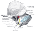
The medial wall and part of the posterior and anterior walls of the right tympanic cavity, lateral view. ("Mastoid antrum" labeled at upper left, in dark circle.)
Details
Identifiers
Latin antrum mastoideum
TA98 A15.3.02.028
TA2 645
FMA 55711
Anatomical terms of bone

The mastoid antrum (tympanic antrum, antrum mastoideum, Valsalva's antrum) is an air space in the petrous portion of the temporal bone, communicating posteriorly with the mastoid cells and anteriorly with the epitympanic recess of the middle ear via the aditus to mastoid antrum (entrance to the mastoid antrum). These air spaces function as sound receptors, provide voice resonance, act as acoustic insulation and dissipation, provide protection from physical damage and reduce the mass of the cranium.[ citation needed ] The roof is formed by the tegmen antri which is a continuation of the tegmen tympani and separates it from the middle cranial fossa. The lateral wall of the antrum is formed by a plate of bone which is an average of 1.5 cm in adults. The mastoid air cell system is a major contributor to middle ear inflammatory diseases. [1]

Contents

Additional images

Related Research Articles

<span class="mw-page-title-main">Facial nerve</span> Cranial nerve VII, for the face and tasting

The facial nerve, also known as the seventh cranial nerve, cranial nerve VII, or simply CN VII, is a cranial nerve that emerges from the pons of the brainstem, controls the muscles of facial expression, and functions in the conveyance of taste sensations from the anterior two-thirds of the tongue. The nerve typically travels from the pons through the facial canal in the temporal bone and exits the skull at the stylomastoid foramen. It arises from the brainstem from an area posterior to the cranial nerve VI and anterior to cranial nerve VIII.

<span class="mw-page-title-main">Glossopharyngeal nerve</span> Cranial nerve IX, for the tongue and pharynx

The glossopharyngeal nerve, also known as the ninth cranial nerve, cranial nerve IX, or simply CN IX, is a cranial nerve that exits the brainstem from the sides of the upper medulla, just anterior to the vagus nerve. Being a mixed nerve (sensorimotor), it carries afferent sensory and efferent motor information. The motor division of the glossopharyngeal nerve is derived from the basal plate of the embryonic medulla oblongata, whereas the sensory division originates from the cranial neural crest.

<span class="mw-page-title-main">Temporal bone</span> Sides and base of skull, connecting to the jaw and occipital bone

The temporal bones are situated at the sides and base of the skull, and lateral to the temporal lobes of the cerebral cortex.

<span class="mw-page-title-main">Ear</span> Organ of hearing and balance

An ear is the organ that enables hearing and body balance using the vestibular system. In mammals the ear is usually described as having three parts: the outer ear, the middle ear and the inner ear. The outer ear consists of the pinna and the ear canal. Since the outer ear is the only visible portion of the ear in most animals, the word "ear" often refers to the external part alone. The middle ear includes the tympanic cavity and the three ossicles. The inner ear sits in the bony labyrinth, and contains structures which are key to several senses: the semicircular canals, which enable balance and eye tracking when moving; the utricle and saccule, which enable balance when stationary; and the cochlea, which enables hearing. The ear is a self cleaning organ through its relationship with earwax and the ear canals. The ears of vertebrates are placed somewhat symmetrically on either side of the head, an arrangement that aids sound localization.

<span class="mw-page-title-main">Mastoiditis</span> Middle ear disease

Mastoiditis is the result of an infection that extends to the air cells of the skull behind the ear. Specifically, it is an inflammation of the mucosal lining of the mastoid antrum and mastoid air cell system inside the mastoid process. The mastoid process is the portion of the temporal bone of the skull that is behind the ear. The mastoid process contains open, air-containing spaces. Mastoiditis is usually caused by untreated acute otitis media and used to be a leading cause of child mortality. With the development of antibiotics, however, mastoiditis has become quite rare in developed countries where surgical treatment is now much less frequent and more conservative, unlike former times.

<span class="mw-page-title-main">Tympanic cavity</span> Small cavity surrounding the bones of the middle ear

The tympanic cavity is a small cavity surrounding the bones of the middle ear. Within it sit the ossicles, three small bones that transmit vibrations used in the detection of sound.

<span class="mw-page-title-main">Stylomastoid artery</span>

The stylomastoid artery enters the stylomastoid foramen and supplies the tympanic cavity, the tympanic antrum and mastoid cells, and the semicircular canals. It is a branch of the posterior auricular artery, and thus part of the external carotid arterial system.

<span class="mw-page-title-main">Jugular fossa</span>

The jugular fossa is a deep depression in the inferior part of the temporal bone at the base of the skull. It lodges the bulb of the internal jugular vein.

<span class="mw-page-title-main">Transverse sinuses</span>

The transverse sinuses, within the human head, are two areas beneath the brain which allow blood to drain from the back of the head. They run laterally in a groove along the interior surface of the occipital bone. They drain from the confluence of sinuses to the sigmoid sinuses, which ultimately connect to the internal jugular vein. See diagram : labeled under the brain as "SIN. TRANS.".

<span class="mw-page-title-main">Posterior auricular artery</span> Small artery

The posterior auricular artery is a small artery that arises from the external carotid artery. It ascends along the side of the head. It supplies several muscles of the neck and several structures of the head.

<span class="mw-page-title-main">Squamous part of temporal bone</span> Front and upper part of the sides of the skull base

The squamous part of temporal bone, or temporal squama, forms the front and upper part of the temporal bone, and is scale-like, thin, and translucent.

<span class="mw-page-title-main">Mastoid part of the temporal bone</span> Back part of the sides of the skull base

The mastoid part of the temporal bone is the posterior (back) part of the temporal bone, one of the bones of the skull. Its rough surface gives attachment to various muscles and it has openings for blood vessels. From its borders, the mastoid part articulates with two other bones.

<span class="mw-page-title-main">Petrous part of the temporal bone</span> Feature at the base of the human skull

The petrous part of the temporal bone is pyramid-shaped and is wedged in at the base of the skull between the sphenoid and occipital bones. Directed medially, forward, and a little upward, it presents a base, an apex, three surfaces, and three angles, and houses in its interior, the components of the inner ear. The petrous portion is among the most basal elements of the skull and forms part of the endocranium. Petrous comes from the Latin word petrosus, meaning "stone-like, hard". It is one of the densest bones in the body. In other mammals, it is a separate bone, the petrosal bone.

<span class="mw-page-title-main">Tympanic part of the temporal bone</span> Middle part of the sides of the skull base, surrounding the ear canal

The tympanic part of the temporal bone is a curved plate of bone lying below the squamous part of the temporal bone, in front of the mastoid process, and surrounding the external part of the ear canal.

<span class="mw-page-title-main">Tympanic nerve</span>

The tympanic nerve is a branch of the glossopharyngeal nerve found near the ear. It gives sensation to the middle ear, the Eustachian tube, the parotid gland, and mastoid air cells. It gives parasympathetic to supply to the parotid gland via the otic ganglion and the auriculotemporal nerve.

<span class="mw-page-title-main">Mastoid cells</span> Air-filled cavities in the temporal bone

The mastoid cells are air-filled cavities within the mastoid process of the temporal bone of the cranium. The mastoid cells are a form of skeletal pneumaticity. Infection in these cells is called mastoiditis.

<span class="mw-page-title-main">Pyramidal eminence</span>

The pyramidal eminence is a hollow conical projection upon the posterior wall of the tympanic cavity of the middle ear. The stapedius muscle arises in the hollow of the eminence and its tendon exits through its apex.

<span class="mw-page-title-main">Aditus to mastoid antrum</span>

The aditus to mastoid antrum, is a large, irregular opening upon the posterior wall of the tympanic cavity by which the mastoid antrum communicates with the epitympanic recess of the tympanic cavity. The walls of the antrum are lined by mucosa which is continuous with that lining the mastoid cells and tympanic cavity.

<span class="mw-page-title-main">Epitympanic recess</span> Hollow located on the superior/roof aspect of the middle ear

The epitympanic recess is the portion of the tympanic cavity situated superior to the tympanic membrane. The recess lodges the head of malleus, and the body of incus.

Koerner's septum is an anatomic boundary in the temporal bone formed by the petrosquamous suture between the petrous and squamosal portions of the mastoid air cells, at the anatomic level of the mastoid antrum. Along with the middle ear ossicles, it is usually eroded in middle ear cholesteatomas. Superiorly, this continues as the petrosquamous suture, a normal anatomic structure that can be mistaken for fractures on temporal bone CT. It is surgically important as it may cause difficulty in locating the antrum and the deeper cells and thus may lead to incomplete removal of disease at mastoidectomy.

References

  1. Koç, Ahmet; Karaaslan, Osman; Koç, Turgay (2004). "Mastoid air cell system" (PDF). Otoscope. 4: 144–54. Archived from the original (PDF) on 2014-05-22. Retrieved 2012-01-15.