In anatomy, the atlas (C1) is the most superior (first) cervical vertebra of the spine and is located in the neck.

The occipital bone is a cranial dermal bone and the main bone of the occiput. It is trapezoidal in shape and curved on itself like a shallow dish. The occipital bone overlies the occipital lobes of the cerebrum. At the base of the skull in the occipital bone, there is a large oval opening called the foramen magnum, which allows the passage of the spinal cord.

The obliquus capitis superior muscle is a small muscle in the upper back part of the neck. It is one of the suboccipital muscles. It attaches inferiorly at the transverse process of the atlas ; it attaches superiorly at the external surface of the occipital bone. The muscle is innervated by the suboccipital nerve.

An occipital bun, also called an occipital spur, occipital knob, chignon hook or inion hook, is a prominent bulge or projection of the occipital bone at the back of the skull. It is important in scientific descriptions of classic Neanderthal crania. It is found among archaic Homo species, as well as Upper Pleistocene Homo sapiens and present-day human populations.

The occipitalis muscle is a muscle which covers parts of the skull. Some sources consider the occipital muscle to be a distinct muscle. However, Terminologia Anatomica currently classifies it as part of the occipitofrontalis muscle along with the frontalis muscle.

The epicranial aponeurosis is an aponeurosis. It covers the upper part of the skull in humans and many other animals.

The rectus capitis posterior major is a muscle in the upper back part of the neck. It is one of the suboccipital muscles. Its inferior attachment is at the spinous process of the axis ; its superior attachment is onto the outer surface of the occipital bone on and around the side part of the inferior nuchal line. The muscle is innervated by the suboccipital nerve. The muscle acts to extend the head and rotate the head to its side.

The rectus capitis posterior minor is a muscle in the upper back part of the neck. It is one of the suboccipital muscles. Its inferior attachment is at the posterior arch of atlas; its superior attachment is onto the occipital bone at and below the inferior nuchal line. The muscle is innervated by the suboccipital nerve. The muscle acts as a weak extensor of the head.

The occipitofrontalis muscle is a muscle which covers parts of the skull. It consists of two parts or bellies: the occipital belly, near the occipital bone, and the frontal belly, near the frontal bone. It is supplied by the supraorbital artery, the supratrochlear artery, and the occipital artery. It is innervated by the facial nerve. In humans, the occipitofrontalis helps to create facial expressions.

The erector spinae or spinal erectors is a set of muscles that straighten and rotate the back. The spinal erectors work together with the glutes to maintain stable posture standing or sitting.

The nuchal ligament is a ligament at the back of the neck that is continuous with the supraspinous ligament.

The nuchal lines are four curved lines on the external surface of the occipital bone:

The transverse sinuses, within the human head, are two areas beneath the brain which allow blood to drain from the back of the head. They run laterally in a groove along the interior surface of the occipital bone. They drain from the confluence of sinuses to the sigmoid sinuses, which ultimately connect to the internal jugular vein. See diagram : labeled under the brain as "SIN. TRANS.".

The occipital vein is a vein of the scalp. It originates from a plexus around the external occipital protuberance and superior nuchal line to the back part of the vertex of the skull. It usually drains into the internal jugular vein, but may also drain into the posterior auricular vein. It drains part of the scalp.

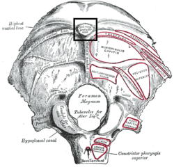
The squamous part of occipital bone is situated above and behind the foramen magnum, and is curved from above downward and from side to side.

Along the internal surface of the occipital bone, at the point of intersection of the four divisions of the cruciform eminence, is the internal occipital protuberance. Running transversely on either side is a groove for the transverse sinus.

The investing layer of deep cervical fascia is the most superficial part of the deep cervical fascia, and encloses the whole neck.

The following outline is provided as an overview of and topical guide to human anatomy:

The groove for transverse sinus is a groove which runs along the internal surface of the occipital bone, running laterally between the superior and inferior fossae of the cruciform eminence. The transverse sinuses travel along this groove.
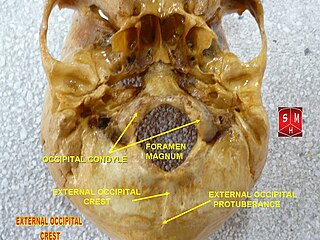
The external occipital crest is part of the external surface of the squamous part of the occipital bone. It is a ridge along the midline, beginning at the external occipital protuberance and descending to the foramen magnum, that gives attachment to the nuchal ligament. It is also called the median nuchal line.
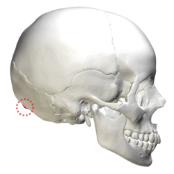
 Position of external occipital protuberance (shown in red). Animation.