Internal occipital crest

Last updated
Internal occipital crest
Internal occipital crest2.png
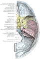
Human skull side view (parietal bones removed). Position of internal occipital crest shown in red.
Gray130 Internal occipital crest.png
Occipital bone. Inner surface. (Position of internal occipital crest labeled as occipital sinus at center.)
Details
Identifiers
Latin crista occipitalis interna
TA A02.1.04.030
FMA 75043
Anatomical terms of bone

In the occipital bone, the lower division of the cruciate eminence is prominent, and is named the internal occipital crest; it bifurcates near the foramen magnum and gives attachment to the falx cerebelli; in the attached margin of this falx is the occipital sinus, which is sometimes duplicated.

Occipital bone saucer-shaped membrane bone situated at the back and lower part of the cranium

The occipital bone is a cranial dermal bone and the main bone of the occiput. It is trapezoidal in shape and curved on itself like a shallow dish. The occipital bone overlies the occipital lobes of the cerebrum. At the base of skull in the occipital bone, there is a large oval opening called the foramen magnum, which allows the passage of the spinal cord.

Foramen magnum Opening in the occipital bone of the cranium

The foramen magnum is a large oval opening (foramen) in the occipital bone of the skull in humans and various other animals. It is one of the several oval or circular openings (foramina) in the base of the skull. The spinal cord, an extension of the medulla, passes through the foramen magnum as it exits the cranial cavity. Apart from the transmission of the medulla oblongata and its membranes, the foramen magnum transmits the vertebral arteries, the anterior and posterior spinal arteries, the tectorial membranes and alar ligaments. It also transmits the spinal component of the accessory nerve into the skull.

Falx cerebelli

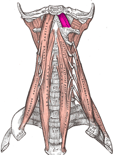
The falx cerebelli is a small sickle shaped fold of dura mater, projecting forwards into the posterior cerebellar notch as well as projecting into the vallecula of the cerebellum between the two cerebellar hemispheres. The name comes from two Latin words: falx, meaning "curved blade or scythe", and cerebellum, meaning "brain". Its base is attached, above, to the under and back part of the tentorium cerebelli; its posterior margin, to the lower division of the vertical crest on the inner surface of the occipital bone. The falx cerebelli generally lies somewhere between 2.8 and 4.5 cm in length and is approximately 1–2 mm thick.

Contents

In the upper part of the internal occipital crest, a small depression is sometimes distinguishable; it is termed the vermian fossa since it is occupied by part of the cerebellar vermis of the cerebellum.

Cerebellar vermis Anatomical structure in the brain

The cerebellar vermis is located in the medial, cortico-nuclear zone of the cerebellum, which resides in the posterior fossa of the cranium. The primary fissure in the vermis curves ventrolaterally to the superior surface of the cerebellum, dividing it into anterior and posterior lobes. Functionally, the vermis is associated with bodily posture and locomotion. The vermis is included within the spinocerebellum and receives somatic sensory input from the head and proximal body parts via ascending spinal pathways.

Cerebellum region of the brain that coordinates motor functions and muscle tone

The cerebellum is a major feature of the hindbrain of all vertebrates. Although usually smaller than the cerebrum, in some animals such as the mormyrid fishes it may be as large as or even larger. In humans, the cerebellum plays an important role in motor control. It may also be involved in some cognitive functions such as attention and language as well as in regulating fear and pleasure responses, but its movement-related functions are the most solidly established. The human cerebellum does not initiate movement, but contributes to coordination, precision, and accurate timing: it receives input from sensory systems of the spinal cord and from other parts of the brain, and integrates these inputs to fine-tune motor activity. Cerebellar damage produces disorders in fine movement, equilibrium, posture, and motor learning in humans.

Additional images

Related Research Articles

Atlas (anatomy) first cervical vertebra of the spine which supports the skull

In anatomy, the atlas (C1) is the most superior (first) cervical vertebra of the spine.

Sphenoid bone unpaired bone situated at the front middle of the skull in front of the temporal bone and basilar part of the occipital bone

The sphenoid bone is an unpaired bone of the neurocranium. It is situated in the middle of the skull towards the front, in front of the temporal bone and the basilar part of the occipital bone. The sphenoid bone is one of the seven bones that articulate to form the orbit. Its shape somewhat resembles that of a butterfly or bat with its wings extended.

Parietal bone bone in the human skull which, when joined together, forms the sides and roof of the cranium

The parietal bones are two bones in the skull which, when joined together at a fibrous joint, form the sides and roof of the cranium. In humans, each bone is roughly quadrilateral in form, and has two surfaces, four borders, and four angles. It is named from the Latin paries (-ietis), wall.

Dura mater Thick membrane surrounding the brain and spinal cord

Dura mater is a thick membrane made of dense irregular connective tissue that surrounds the brain and spinal cord. It is the outermost of the three layers of membrane called the meninges that protect the central nervous system. The other two meningeal layers are the arachnoid mater and the pia mater. The dura surrounds the brain and the spinal cord and is responsible for keeping in the cerebrospinal fluid. It is derived from mesoderm.

Rectus capitis anterior muscle

The rectus capitis anterior is a short, flat muscle, situated immediately behind the upper part of the Longus capitis.

Rectus capitis posterior minor muscle

The rectus capitis posterior minor arises by a narrow pointed tendon from the tubercle on the posterior arch of the atlas, and, widening as it ascends, is inserted into the medial part of the inferior nuchal line of the occipital bone and the surface between it and the foramen magnum, and also takes some attachment to the spinal dura mater.

Occipital sinus

The occipital sinus is the smallest of the dural venous sinuses.

Nuchal lines

The nuchal lines are four curved lines on the external surface of the occipital bone:

Transverse sinuses

The transverse sinuses, within the human head, are two areas beneath the brain which allow blood to drain from the back of the head. They run laterally in a groove along the interior surface of the occipital bone. They drain from the confluence of sinuses to the sigmoid sinuses, which ultimately connect to the internal jugular vein. See diagram : labeled under the brain as "SIN. TRANS.".

Petrous part of the temporal bone

The petrous part of the temporal bone is pyramid-shaped and is wedged in at the base of the skull between the sphenoid and occipital bones. Directed medially, forward, and a little upward, it presents a base, an apex, three surfaces, and three angles, and houses in its interior, the components of the inner ear. The petrous portion is among the most basal elements of the skull and forms part of the endocranium. Petrous comes from the Latin word petrosus, meaning "stone-like, hard". It is one of the densest bones in the body.

Basilar part of occipital bone

The basilar part of the occipital bone extends forward and upward from the foramen magnum, and presents in front an area more or less quadrilateral in outline.

Lateral parts of occipital bone

The lateral parts of the occipital bone are situated at the sides of the foramen magnum; on their under surfaces are the condyles for articulation with the superior facets of the atlas.

Squamous part of occipital bone

The squamous part of occipital bone, is situated above and behind the foramen magnum, and is curved from above downward and from side to side.

Squamous part of the frontal bone

There are two surfaces of the squamous part of the frontal bone: the external surface, and the internal surface.

Anterior cranial fossa houses the projecting frontal lobes of the brain

The anterior cranial fossa is a depression in the floor of the cranial base which houses the projecting frontal lobes of the brain. It is formed by the orbital plates of the frontal, the cribriform plate of the ethmoid, and the small wings and front part of the body of the sphenoid; it is limited behind by the posterior borders of the small wings of the sphenoid and by the anterior margin of the chiasmatic groove. The lesser wings of the sphenoid separate the anterior and middle fossae.

Base of skull inferior area of the skull, composed of the endocranium and lower parts of the skull roof

The base of skull, also known as the cranial base or the cranial floor, is the most inferior area of the skull. It is composed of the endocranium and the lower parts of the skull roof.

Endocranium The lower and inner parts of the skull

The endocranium in comparative anatomy is a part of the skull base in vertebrates and it represents the basal, inner part of the cranium. The term is also applied to the outer layer of the dura mater in human anatomy.

References

This article incorporates text in the public domain from page 131 of the 20th edition of Gray's Anatomy (1918)

The public domain consists of all the creative work to which no exclusive intellectual property rights apply. Those rights may have expired, been forfeited, expressly waived, or may be inapplicable.

<i>Grays Anatomy</i> textbook of human anatomy

Gray's Anatomy is an English language textbook of human anatomy originally written by Henry Gray and illustrated by Henry Vandyke Carter. Earlier editions were called Anatomy: Descriptive and Surgical, Anatomy of the Human Body and Gray's Anatomy: Descriptive and Applied, but the book's name is commonly shortened to, and later editions are titled, Gray's Anatomy. The book is widely regarded as an extremely influential work on the subject, and has continued to be revised and republished from its initial publication in 1858 to the present day. The latest edition of the book, the 41st, was published in September 2015.