Orbital part of frontal bone

Last updated
Orbital part of frontal bone
Gray134.png
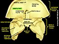
Frontal bone. Outer surface. (The Pars orbitalis is the bottom third.)
Gray135.png
Frontal bone. Inner surface. (The Pars orbitalis is the bottom third.)
Details
Identifiers
Latin pars orbitalis ossis frontalis
TA98 A02.1.03.022
TA2 541
FMA 52849
Anatomical terminology

The orbital or horizontal part of the frontal bone (pars orbitalis) consists of two thin triangular plates, the orbital plates, which form the vaults of the orbits, and are separated from one another by a median gap, the ethmoidal notch.

Contents

Surfaces

Additional images

Related Research Articles

Maxilla

The maxilla in vertebrates is the upper fixed bone of the jaw formed from the fusion of two maxillary bones. In humans, the upper jaw includes the hard palate in the front of the mouth. The two maxillary bones are fused at the intermaxillary suture, forming the anterior nasal spine. This is similar to the mandible, which is also a fusion of two mandibular bones at the mandibular symphysis. The mandible is the movable part of the jaw.

Ethmoid bone

The ethmoid bone is an unpaired bone in the skull that separates the nasal cavity from the brain. It is located at the roof of the nose, between the two orbits. The cubical bone is lightweight due to a spongy construction. The ethmoid bone is one of the bones that make up the orbit of the eye.

Lacrimal bone

The lacrimal bone is a small and fragile bone of the facial skeleton; it is roughly the size of the little fingernail. It is situated at the front part of the medial wall of the orbit. It has two surfaces and four borders. Several bony landmarks of the lacrimal bone function in the process of lacrimation or crying. Specifically, the lacrimal bone helps form the nasolacrimal canal necessary for tear translocation. A depression on the anterior inferior portion of the bone, the lacrimal fossa, houses the membranous lacrimal sac. Tears or lacrimal fluid, from the lacrimal glands, collect in this sac during excessive lacrimation. The fluid then flows through the nasolacrimal duct and into the nasopharynx. This drainage results in what is commonly referred to a runny nose during excessive crying or tear production. Injury or fracture of the lacrimal bone can result in posttraumatic obstruction of the lacrimal pathways.

Inferior nasal concha

The inferior nasal concha is one of the three paired nasal conchae in the nose. It extends horizontally along the lateral wall of the nasal cavity and consists of a lamina of spongy bone, curled upon itself like a scroll,. The inferior nasal conchae are considered a pair of facial bones. As the air passes through the turbinates, the air is churned against these mucosa-lined bones in order to receive warmth, moisture and cleansing. Superior to inferior nasal concha are the middle nasal concha and superior nasal concha which arise from the cranial portion of the skull. Hence, these two are considered as a part of the cranial bones.

Orbit (anatomy) Cavity or socket of the skull in which the eye and its appendages are situated

In anatomy, the orbit is the cavity or socket of the skull in which the eye and its appendages are situated. "Orbit" can refer to the bony socket, or it can also be used to imply the contents. In the adult human, the volume of the orbit is 30 millilitres, of which the eye occupies 6.5 ml. The orbital contents comprise the eye, the orbital and retrobulbar fascia, extraocular muscles, cranial nerves II, III, IV, V, and VI, blood vessels, fat, the lacrimal gland with its sac and duct, the eyelids, medial and lateral palpebral ligaments, check ligaments, the suspensory ligament, septum, ciliary ganglion and short ciliary nerves.

Cribriform plate Part of the ethmoid bone that transmits olfactory nerves between the nasal cavity and the neurocranium

In mammalian anatomy, the cribriform plate, horizontal lamina or lamina cribrosa is part of the ethmoid bone. It is received into the ethmoidal notch of the frontal bone and roofs in the nasal cavities. It supports the olfactory bulb, and is perforated by olfactory foramina for the passage of the olfactory nerves to the roof of the nasal cavity to convey smell to the brain. The foramina at the medial part of the groove allow the passage of the nerves to the upper part of the nasal septum while the foramina at the lateral part transmit the nerves to the superior nasal concha.

Maxillary sinus Largest of the paranasal sinuses, and drains into the middle meatus of the nose

The pyramid-shaped maxillary sinus is the largest of the paranasal sinuses, and drains into the middle meatus of the nose through the osteomeatal complex.

Ethmoid sinus

The ethmoid sinuses or ethmoid air cells of the ethmoid bone are one of the four paired paranasal sinuses. The cells are variable in both size and number in the lateral mass of each of the ethmoid bones and cannot be palpated during an extraoral examination. They are divided into anterior and posterior groups. The ethmoid air cells are numerous thin-walled cavities situated in the ethmoidal labyrinth and completed by the frontal, maxilla, lacrimal, sphenoidal, and palatine bones. They lie between the upper parts of the nasal cavities and the orbits, and are separated from these cavities by thin bony lamellae.

Ophthalmic nerve

The ophthalmic nerve (V1) is one of three divisions of the trigeminal nerve (CN V). It has three branches that provide sensory innervation to the eye, skin of the upper face and anterior scalp.

Nasociliary nerve

The nasociliary nerve is a branch of the ophthalmic nerve, itself a branch of the trigeminal nerve. It is intermediate in size between the other two branches of the ophthalmic nerve, the frontal nerve and lacrimal nerve.

Greater wing of sphenoid bone

The greater wing of the sphenoid bone, or alisphenoid, is a bony process of the sphenoid bone; there is one on each side, extending from the side of the body of the sphenoid and curving upward, laterally, and backward.

Ethmoidal labyrinth

The ethmoidal labyrinth or lateral mass of the ethmoid bone consists of a number of thin-walled cellular cavities, the ethmoid air cells, arranged in three groups, anterior, middle, and posterior, and interposed between two vertical plates of bone; the lateral plate forms part of the orbit, the medial plate forms part of the nasal cavity. In the disarticulated bone many of these cells are opened into, but when the bones are articulated, they are closed in at every part, except where they open into the nasal cavity.

Anterior ethmoidal artery

The anterior ethmoidal artery, is a branch of the ophthalmic artery in the orbit. It exits the orbit through the anterior ethmoidal foramen. The posterior ethmoidal artery is posterior to it.

Squamous part of the frontal bone

There are two surfaces of the squamous part of the frontal bone: the external surface, and the internal surface.

Middle cranial fossa

The middle cranial fossa, deeper than the anterior cranial fossa, is narrow medially and widens laterally to the sides of the skull. It is separated from the posterior fossa by the clivus and the petrous crest.

Anterior cranial fossa

The anterior cranial fossa is a depression in the floor of the cranial base which houses the projecting frontal lobes of the brain. It is formed by the orbital plates of the frontal, the cribriform plate of the ethmoid, and the small wings and front part of the body of the sphenoid; it is limited behind by the posterior borders of the small wings of the sphenoid and by the anterior margin of the chiasmatic groove. The lesser wings of the sphenoid separate the anterior and middle fossae.

Perpendicular plate of palatine bone

The perpendicular plate of palatine bone is the vertical part of the palatine bone, and is thin, of an oblong form, and presents two surfaces and four borders.

Frontal process of maxilla

The frontal process of maxilla is a strong plate, which projects upward, medialward, and backward from the maxilla, forming part of the lateral boundary of the nose.

Body of sphenoid bone

The body of the sphenoid bone, more or less cubical in shape, is hollowed out in its interior to form two large cavities, the sphenoidal sinuses, which are separated from each other by a septum.

Outline of human anatomy Overview of and topical guide to human anatomy

The following outline is provided as an overview of and topical guide to human anatomy:

References

PD-icon.svgThis article incorporates text in the public domain from page 137 of the 20th edition of Gray's Anatomy (1918)