
The occipital bone is a cranial dermal bone and the main bone of the occiput. It is trapezoidal in shape and curved on itself like a shallow dish. The occipital bone overlies the occipital lobes of the cerebrum. At the base of the skull in the occipital bone, there is a large oval opening called the foramen magnum, which allows the passage of the spinal cord.

The posterior cranial fossa is the part of the cranial cavity located between the foramen magnum, and tentorium cerebelli. It is formed by the sphenoid bones, temporal bones, and occipital bone. It lodges the cerebellum, and parts of the brainstem.

In anatomy, a process is a projection or outgrowth of tissue from a larger body. For instance, in a vertebra, a process may serve for muscle attachment and leverage, or to fit, with another vertebra. The word is also used at the microanatomic level, where cells can have processes such as cilia or pedicels. Depending on the tissue, processes may also be called by other terms, such as apophysis, tubercle, or protuberance.

The cerebellar tentorium or tentorium cerebelli is an extension of the dura mater between the inferior aspect of the occipital lobes and the superior aspect of the cerebellum. The free border of the tentorium gives passage to the midbrain.
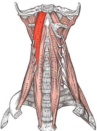
The longus capitis muscle, is broad and thick above, narrow below, and arises by four tendinous slips, from the anterior tubercles of the transverse processes of the third, fourth, fifth, and sixth cervical vertebræ, and ascends, converging toward its fellow of the opposite side, to be inserted into the inferior surface of the basilar part of the occipital bone.

The confluence of sinuses, torcular Herophili, or torcula is the connecting point of the superior sagittal sinus, straight sinus, and occipital sinus. It is below the internal occipital protuberance of the skull. It drains venous blood from the brain into the transverse sinuses. It may be affected by arteriovenous fistulas, a thrombus, major trauma, or surgical damage, and may be imaged with many radiology techniques.
The superior sagittal sinus, within the human head, is an unpaired area along the attached margin of the falx cerebri. It allows blood to drain from the lateral aspects of anterior cerebral hemispheres to the confluence of sinuses. Cerebrospinal fluid drains through arachnoid granulations into the superior sagittal sinus and is returned to venous circulation.

The occipital artery is a branch of the external carotid artery that provides arterial supply to the back of the scalp, sternocleidomastoid muscles, and deep muscles of the back and neck.

The erector spinae or spinal erectors is a set of muscles that straighten and rotate the back. The spinal erectors work together with the glutes to maintain stable posture standing or sitting.

The nuchal lines are four curved lines on the external surface of the occipital bone:
The transverse sinuses, within the human head, are two areas beneath the brain which allow blood to drain from the back of the head. They run laterally in a groove along the interior surface of the occipital bone. They drain from the confluence of sinuses to the sigmoid sinuses, which ultimately connect to the internal jugular vein. See diagram : labeled under the brain as "SIN. TRANS.".

The occipital vein is a vein of the scalp. It originates from a plexus around the external occipital protuberance and superior nuchal line to the back part of the vertex of the skull. It usually drains into the internal jugular vein, but may also drain into the posterior auricular vein. It drains part of the scalp.

The mastoid part of the temporal bone is the posterior (back) part of the temporal bone, one of the bones of the skull. Its rough surface gives attachment to various muscles and it has openings for blood vessels. From its borders, the mastoid part articulates with two other bones.

The squamous part of occipital bone is situated above and behind the foramen magnum, and is curved from above downward and from side to side.
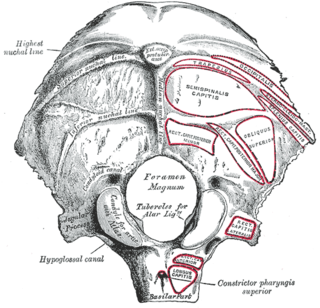
The jugular tubercle is a rounded prominence/oval elevation upon the superior surface of the occipital condyle at the junction of the basilar part and lateral part of the occipital bone, just medial to the jugular foramen on either side of the foramen magnum.

Near the middle of the squamous part of occipital bone is the external occipital protuberance, the highest point of which is referred to as the inion. The inion is the most prominent projection of the protuberance which is located at the posterioinferior part of the human skull. The nuchal ligament and trapezius muscle attach to it.

The occipital condyles are undersurface protuberances of the occipital bone in vertebrates, which function in articulation with the superior facets of the atlas vertebra.
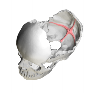
The cruciform eminence divides the deeply concave internal surface of the occipital bone into four fossae:

The following outline is provided as an overview of and topical guide to human anatomy:

The groove for transverse sinus is a groove which runs along the internal surface of the occipital bone, running laterally between the superior and inferior fossae of the cruciform eminence. The transverse sinuses travel along this groove.
 Human skull. Internal occipital protuberance shown in red. Parietal bones and temporal bones are removed.

