Diaphragma sellae

Last updated
Diaphragma sellae
Gray766.png
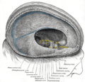
Tentorium cerebelli seen from above. (Diaphragma sellae labeled at upper left.)
Details
Identifiers
Latin diaphragma sellae
TA98 A14.1.01.107
TA2 5378
FMA 78540
Anatomical terminology

The diaphragma sellae or sellar diaphragm is a small, circular sheet of dura mater forming an (incomplete) roof over the sella turcica and covering the pituitary gland lodged therein. The diaphragma sellae forms a central opening to accommodate the passage of the pituitary stalk (infundibulum) [1] which interconnects the pituitary gland and the hypothalamus.

Contents

The diaphragma sellae is an important neurosurgical landmark. [1]

Anatomy

Boundaries

The diaphragma sellae has a posterior boundary at the dorsum sellae and an anterior boundary at the tuberculum sellae along with the two small eminences (one on either side) called the middle clinoid processes.

Variation

The opening formed by the diaphragma sellae varies greatly in size between individuals. [1]

Clinical significance

Pituitary tumours may grow to extend superiorly beyond the diaphragma sellae. [1] Violation of the diaphragma sellae during an endoscopic endonasal transsphenoidal pituitary tumor resection will result in a cerebrospinal fluid leak.[ citation needed ]

Additional images

References

  1. 1 2 3 4 Standring, Susan (2020). Gray's Anatomy: The Anatomical Basis of Clinical Practice (42nd ed.). New York. p. 398. ISBN   978-0-7020-7707-4. OCLC   1201341621.{{cite book}}: CS1 maint: location missing publisher (link)

PD-icon.svgThis article incorporates text in the public domain from page 814 of the 20th edition of Gray's Anatomy (1918)