Choroid plexus

Last updated
Choroid plexus
Gray708.png
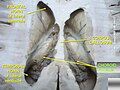
Choroid plexus shown in the fourth ventricle
Gray723.png
Details
Identifiers
Latin plexus choroideus
MeSH D002831
NeuroNames 1377
TA98 A14.1.09.279
A14.1.01.307
A14.1.01.306
A14.1.01.304
A14.1.05.715
TA2 5654, 5786, 5980
FMA 61934
Anatomical terms of neuroanatomy

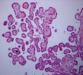
The choroid plexus, or plica choroidea, is a plexus of cells that arises from the tela choroidea in each of the ventricles of the brain. [1] Regions of the choroid plexus produce and secrete most of the cerebrospinal fluid (CSF) of the central nervous system. [2] [3] The choroid plexus consists of modified ependymal cells surrounding a core of capillaries and loose connective tissue. [3] Multiple cilia on the ependymal cells move to circulate the cerebrospinal fluid. [4]

Contents

Structure

Location

Scheme of roof of fourth ventricle. The arrow is in the median aperture.
1: Inferior medullary velum
2: Choroid plexus
3: Cisterna magna of subarachnoid space
4: Central canal
5: Corpora quadrigemina
6: Cerebral peduncle
7: Superior medullary velum
8: Ependymal lining of ventricle
9: Pontine cistern of subarachnoid space Gray708.svg
Scheme of roof of fourth ventricle. The arrow is in the median aperture.
1: Inferior medullary velum
2: Choroid plexus
3: Cisterna magna of subarachnoid space
4: Central canal
5: Corpora quadrigemina
6: Cerebral peduncle
7: Superior medullary velum
8: Ependymal lining of ventricle
9: Pontine cistern of subarachnoid space

There is a choroid plexus in each of the four ventricles. In the lateral ventricles, it is found in the body, and continued in an enlarged amount in the atrium. There is no choroid plexus in the anterior horn. In the third ventricle, there is a small amount in the roof that is continuous with that in the body, via the interventricular foramina, the channels that connect the lateral ventricles with the third ventricle. A choroid plexus is in part of the roof of the fourth ventricle.

Microanatomy

The choroid plexus consists of a layer of cuboidal epithelial cells surrounding a core of capillaries and loose connective tissue. [3] The epithelium of the choroid plexus is continuous with the ependymal cell layer (ventricular layer) that lines the ventricular system. [5] Progenitor ependymal cells are monociliated but they differentiate into multiciliated ependymal cells. [6] [7] Unlike the ependyma, the choroid plexus epithelial layer has tight junctions [8] between the cells on the side facing the ventricle (apical surface). These tight junctions prevent the majority of substances from crossing the cell layer into the cerebrospinal fluid (CSF); thus the choroid plexus acts as a blood–CSF barrier. The choroid plexus folds into many villi around each capillary, creating frond-like processes that project into the ventricles. The villi, along with a brush border of microvilli, greatly increase the surface area of the choroid plexus.[ citation needed ] CSF is formed as plasma is filtered from the blood through the epithelial cells. Choroid plexus epithelial cells actively transport sodium ions into the ventricles and water follows the resulting osmotic gradient. [9]

The choroid plexus consists of many capillaries, separated from the ventricles by choroid epithelial cells. Fluid filters through these cells from blood to become cerebrospinal fluid. There is also much active transport of substances into, and out of, the CSF as it is made.

Function

CSF circulation CSF circulation.png
CSF circulation

The choroid plexus regulates the production and composition of cerebrospinal fluid (CSF), that provides the protective buoyancy for the brain. [2] [10] CSF acts as a medium for the glymphatic filtration system that facilitates the removal of metabolic waste from the brain, and the exchange of biomolecules and xenobiotics into and out of the brain. [10] [11] In this way the choroid plexus has a very important role in helping to maintain the delicate extracellular environment required by the brain to function optimally.

The choroid plexus is also a major source of transferrin secretion that plays a part in iron homeostasis in the brain. [12] [13]

Blood–cerebrospinal fluid barrier

The blood–cerebrospinal fluid barrier (BCSFB) is a fluid–brain barrier that is composed of a pair of membranes that separate blood from CSF at the capillary level and CSF from brain tissue. [14] The blood–CSF boundary at the choroid plexus is a membrane composed of epithelial cells and tight junctions that link them. [14] There is a CSF-brain barrier at the level of the pia mater, but only in the embryo. [15]

Similar to the blood–brain barrier, the blood–CSF barrier functions to prevent the passage of most blood-borne substances into the brain, while selectively permitting the passage of specific substances (such as nutrients) into the brain and facilitating the removal of brain metabolites and metabolic products into the blood. [14] [16] Despite the similar function between the BBB and BCSFB, each facilitates the transport of different substances into the brain due to the distinctive structural characteristics of each of the two barrier systems. [14] For a number of substances, the BCSFB is the primary site of entry into brain tissue. [14]

The blood–cerebrospinal fluid barrier has also been shown to modulate the entry of leukocytes from the blood to the central nervous system. The choroid plexus cells secrete cytokines that recruit monocyte-derived macrophages, among other cells, to the brain. This cellular trafficking has implications both in normal brain homeostasis and in neuroinflammatory processes. [17]

Clinical significance

Choroid plexus cysts

During fetal development, some choroid plexus cysts may form. These fluid-filled cysts can be detected by a detailed second trimester ultrasound. The finding is relatively common, with a prevalence of ~1%. Choroid plexus cysts are usually an isolated finding. [18] The cysts typically disappear later during pregnancy, and are usually harmless. They have no effect on infant and early childhood development. [19]

Choroid plexus cysts are associated with a 1% risk of fetal aneuploidy. [20] The risk of aneuploidy increases to 10.5-12% if other risk factors or ultrasound findings are noted. Size, location, disappearance or progression, and whether the cysts are found on both sides or not do not affect the risk of aneuploidy. 44-50% of Edwards syndrome (trisomy 18) cases will present with choroid plexus cysts, as well 1.4% of Down syndrome (trisomy 21) cases. ~75% of abnormal karyotypes associated with choroid plexus cysts are trisomy 18, while the remainder are trisomy 21. [18]

Other

There are three graded types of choroid plexus tumor that mainly affect young children. These types of cancer are rare.

Etymology

Choroid plexus translates from the Latin plexus chorioides, [21] which mirrors Ancient Greek χοριοειδές πλέγμα. [22] The word chorion was used by Galen to refer to the outer membrane enclosing the fetus. Both meanings of the word plexus are given as pleating, or braiding. [22] As often happens language changes and the use of both choroid or chorioid is both accepted. Nomina Anatomica (now Terminologia Anatomica) reflected this dual usage.

Additional images

See also

Related Research Articles

<span class="mw-page-title-main">Cerebrospinal fluid</span> Clear, colorless bodily fluid found in the brain and spinal cord

Cerebrospinal fluid (CSF) is a clear, colorless body fluid found within the tissue that surrounds the brain and spinal cord of all vertebrates.

<span class="mw-page-title-main">Blood–brain barrier</span> Semipermeable capillary border that allows selective passage of blood constituents into the brain

The blood–brain barrier (BBB) is a highly selective semipermeable border of endothelial cells that regulates the transfer of solutes and chemicals between the circulatory system and the central nervous system, thus protecting the brain from harmful or unwanted substances in the blood. The blood–brain barrier is formed by endothelial cells of the capillary wall, astrocyte end-feet ensheathing the capillary, and pericytes embedded in the capillary basement membrane. This system allows the passage of some small molecules by passive diffusion, as well as the selective and active transport of various nutrients, ions, organic anions, and macromolecules such as glucose and amino acids that are crucial to neural function.

<span class="mw-page-title-main">Ventricular system</span> Set of structures containing cerebrospinal fluid in the brain

In neuroanatomy, the ventricular system is a set of four interconnected cavities known as cerebral ventricles in the brain. Within each ventricle is a region of choroid plexus which produces the circulating cerebrospinal fluid (CSF). The ventricular system is continuous with the central canal of the spinal cord from the fourth ventricle, allowing for the flow of CSF to circulate.

<span class="mw-page-title-main">Pia mater</span> Delicate innermost layer of the meninges, the membranes surrounding the brain and spinal cord

Pia mater, often referred to as simply the pia, is the delicate innermost layer of the meninges, the membranes surrounding the brain and spinal cord. Pia mater is medieval Latin meaning "tender mother". The other two meningeal membranes are the dura mater and the arachnoid mater. Both the pia and arachnoid mater are derivatives of the neural crest while the dura is derived from embryonic mesoderm. The pia mater is a thin fibrous tissue that is permeable to water and small solutes. The pia mater allows blood vessels to pass through and nourish the brain. The perivascular space between blood vessels and pia mater is proposed to be part of a pseudolymphatic system for the brain. When the pia mater becomes irritated and inflamed the result is meningitis.

<span class="mw-page-title-main">Ependyma</span>

The ependyma is the thin neuroepithelial lining of the ventricular system of the brain and the central canal of the spinal cord. The ependyma is one of the four types of neuroglia in the central nervous system (CNS). It is involved in the production of cerebrospinal fluid (CSF), and is shown to serve as a reservoir for neuroregeneration.

<span class="mw-page-title-main">Choroid plexus cyst</span> Medical condition

Choroid plexus cysts (CPCs) are cysts that occur within choroid plexus of the brain. They are the most common type of intraventricular cyst, occurring in 1% of all pregnancies.

<span class="mw-page-title-main">Median aperture</span>

The median aperture is an opening of the fourth ventricle at the caudal portion of the roof of the fourth ventricle. It allows flow of cerebrospinal fluid (CSF) from the fourth ventricle into the cisterna magna. The other two openings of the fourth ventricle are the lateral apertures - one on either side. Nonetheless, the median aperture accounts for most of the outflow of CSF out of the fourth ventricle. The median aperture varies in size.

<span class="mw-page-title-main">Lateral ventricles</span> Two largest ventricles in each cerebral hemisphere

The lateral ventricles are the two largest ventricles of the brain and contain cerebrospinal fluid (CSF). Each cerebral hemisphere contains a lateral ventricle, known as the left or right lateral ventricle, respectively.

<span class="mw-page-title-main">Interventricular foramina (neuroanatomy)</span> It is part of diencephalon that makes connection between lateral and third ventricular

In the brain, the interventricular foramina are channels that connect the paired lateral ventricles with the third ventricle at the midline of the brain. As channels, they allow cerebrospinal fluid (CSF) produced in the lateral ventricles to reach the third ventricle and then the rest of the brain's ventricular system. The walls of the interventricular foramina also contain choroid plexus, a specialized CSF-producing structure, that is continuous with that of the lateral and third ventricles above and below it.

<span class="mw-page-title-main">Central canal</span> Cerebrospinal fluid-filled space around the spinal cord

The central canal is the cerebrospinal fluid-filled space that runs through the spinal cord. The central canal lies below and is connected to the ventricular system of the brain, from which it receives cerebrospinal fluid, and shares the same ependymal lining. The central canal helps to transport nutrients to the spinal cord as well as protect it by cushioning the impact of a force when the spine is affected.

<span class="mw-page-title-main">Circumventricular organs</span> Interfaces between the brain and the circulatory system

Circumventricular organs (CVOs) are structures in the brain characterized by their extensive and highly permeable capillaries, unlike those in the rest of the brain where there exists a blood–brain barrier (BBB) at the capillary level. Although the term "circumventricular organs" was originally proposed in 1958 by Austrian anatomist Helmut O. Hofer concerning structures around the brain ventricular system, the penetration of blood-borne dyes into small specific CVO regions was discovered in the early 20th century. The permeable CVOs enabling rapid neurohumoral exchange include the subfornical organ (SFO), the area postrema (AP), the vascular organ of lamina terminalis, the median eminence, the pituitary neural lobe, and the pineal gland.

<span class="mw-page-title-main">Tela choroidea</span>

The tela choroidea is a region of meningeal pia mater that adheres to the underlying ependyma, and gives rise to the choroid plexus in each of the brain’s four ventricles. Tela is Latin for woven and is used to describe a web-like membrane or layer. The tela choroidea is a very thin part of the loose connective tissue of pia mater overlying and closely adhering to the ependyma. It has a rich blood supply. The ependyma and vascular pia mater – the tela choroidea, form regions of minute projections known as a choroid plexus that projects into each ventricle. The choroid plexus produces most of the cerebrospinal fluid of the central nervous system that circulates through the ventricles of the brain, the central canal of the spinal cord, and the subarachnoid space. The tela choroidea in the ventricles forms from different parts of the roof plate in the development of the embryo.

<span class="mw-page-title-main">Superior medullary velum</span> Thin layer between the superior cerebellar peduncles

The superior medullary velum is a thin, transparent lamina of white matter which - together with the inferior medullary velum - forms the roof of the fourth ventricle. It extends between the two superior cerebellar peduncles. The lingula of cerebellum covers - and adheres to - its dorsal surface.

<span class="mw-page-title-main">Subcommissural organ</span>

The subcommissural organ (SCO) is one of the circumventricular organs of the brain. It is a small glandular structure that is located in the posterior region of the third ventricle, near the entrance of the cerebral aqueduct.

<span class="mw-page-title-main">Tanycyte</span>

Tanycytes are special ependymal cells found in the third ventricle of the brain, and on the floor of the fourth ventricle and have processes extending deep into the hypothalamus. It is possible that their function is to transfer chemical signals from the cerebrospinal fluid to the central nervous system.

<span class="mw-page-title-main">Choroid plexus carcinoma</span> Medical condition

A choroid plexus carcinoma is a type of choroid plexus tumor that affects the choroid plexus of the brain. It is considered the worst of the three grades of chord plexus tumors, having a much poorer prognosis than choroid atypical plexus papilloma and choroid plexus papilloma. The disease creates lesions in the brain and increases cerebrospinal fluid volume, resulting in hydrocephalus.

<span class="mw-page-title-main">Central nervous system cyst</span> Medical condition

A central nervous system cyst is a type of cyst that presents and affects part of the central nervous system (CNS). They are usually benign and filled with either cerebrospinal fluid, blood, or tumor cells. CNS cysts are classified into two categories: cysts that originate from non-central nervous system tissue, migrate to, and form on a portion of the CNS, and cysts that originate within central nervous system tissue itself. Within these two categories, there are many types of CNS cysts that have been identified from previous studies.

Bobble-head doll syndrome is a rare neurological movement disorder in which patients, usually children around age 3, begin to bob their head and shoulders forward and back, or sometimes side-to-side, involuntarily, in a manner reminiscent of a bobblehead doll. The syndrome is related to cystic lesions and swelling of the third ventricle in the brain.

<span class="mw-page-title-main">Glymphatic system</span> System for waste clearance in the central nervous system of vertebrates

The glymphatic system is a system for waste clearance in the central nervous system (CNS) of vertebrates. According to this model, cerebrospinal fluid (CSF) flows into the paravascular space around cerebral arteries, combining with interstitial fluid (ISF) and parenchymal solutes, and exiting down venous paravascular spaces. The pathway consists of a para-arterial influx route for CSF to enter the brain parenchyma, coupled to a clearance mechanism for the removal of interstitial fluid (ISF) and extracellular solutes from the interstitial compartments of the brain and spinal cord. Exchange of solutes between CSF and ISF is driven primarily by arterial pulsation and regulated during sleep by the expansion and contraction of brain extracellular space. Clearance of soluble proteins, waste products, and excess extracellular fluid is accomplished through convective bulk flow of ISF, facilitated by astrocytic aquaporin 4 (AQP4) water channels.

<span class="mw-page-title-main">Intracerebroventricular injection</span> Injection into the cerebrospinal fluid

Intracerebroventricular injection is a route of administration for drugs via injection into the cerebral ventricles so that it reaches the cerebrospinal fluid (CSF). This route of administration is often used to bypass the blood-brain barrier because it can prevent important medications from reaching the central nervous system. This injection method is widely used in diseased mice models to study the effect of drugs, plasmid DNA, and viral vectors on the central nervous system. In humans, ICV injection can be used for the administration of drugs for various reasons. Examples include the treatment of Spinal Muscular Atrophy (SMA), the administration of chemotherapy in gliomas, and the administration of drugs for long-term pain management. ICV injection is also used in the creation of diseased animal models specifically to model neurological disorders.

References

PD-icon.svgThis article incorporates text in the public domain from page 798 of  page 841 of  page 816 of the 20th edition of Gray's Anatomy (1918)

  1. Sadler, T. (2010). Langman's medical embryology (11th ed.). Philadelphia: Lippincott William & Wilkins. p. 305. ISBN   978-0-7817-9069-7.
  2. 1 2 Damkier, HH; Brown, PD; Praetorius, J (October 2013). "Cerebrospinal fluid secretion by the choroid plexus" (PDF). Physiological Reviews. 93 (4): 1847–92. doi:10.1152/physrev.00004.2013. PMID   24137023. S2CID   11473603.
  3. 1 2 3 Lun, MP; Monuki, ES; Lehtinen, MK (August 2015). "Development and functions of the choroid plexus-cerebrospinal fluid system". Nature Reviews. Neuroscience. 16 (8): 445–57. doi:10.1038/nrn3921. PMC   4629451 . PMID   26174708.
  4. Takeda, S; Narita, K (February 2012). "Structure and function of vertebrate cilia, towards a new taxonomy". Differentiation; Research in Biological Diversity. 83 (2): S4-11. doi:10.1016/j.diff.2011.11.002. PMID   22118931.
  5. Javed K, Reddy V, Lui F (1 January 2022). Neuroanatomy, Choroid Plexus. StatPearls. PMID   30844183.
  6. Delgehyr, N; Meunier, A; Faucourt, M; Bosch Grau, M; Strehl, L; Janke, C; Spassky, N (2015). Ependymal cell differentiation, from monociliated to multiciliated cells. Methods in Cell Biology. Vol. 127. pp. 19–35. doi:10.1016/bs.mcb.2015.01.004. ISBN   9780128024515. PMID   25837384.
  7. van Leeuwen LM, Evans RJ, Jim KK, Verboom T, Fang X, Bojarczuk A, Malicki J, Johnston SA, van der Sar AM (February 2018). "A transgenic zebrafish model for the in vivo study of the blood and choroid plexus brain barriers using claudin 5". Biology Open. 7 (2): bio030494. doi:10.1242/bio.030494. PMC   5861362 . PMID   29437557.
  8. Hall, John (2011). Guyton and Hall textbook of medical physiology (12th ed.). Philadelphia, Pa.: Saunders/Elsevier. p. 749. ISBN   978-1-4160-4574-8.
  9. Guyton AC, Hall JE (2005). Textbook of medical physiology (11th ed.). Philadelphia: W.B. Saunders. pp. 764–7. ISBN   978-0-7216-0240-0.
  10. 1 2 Plog BA, Nedergaard M (January 2018). "The Glymphatic System in Central Nervous System Health and Disease: Past, Present, and Future". Annual Review of Pathology. 13: 379–394. doi:10.1146/annurev-pathol-051217-111018. PMC   5803388 . PMID   29195051.
  11. Abbott NJ, Pizzo ME, Preston JE, Janigro D, Thorne RG (March 2018). "The role of brain barriers in fluid movement in the CNS: is there a 'glymphatic' system?". Acta Neuropathologica. 135 (3): 387–407. doi: 10.1007/s00401-018-1812-4 . PMID   29428972.
  12. Moos, T (November 2002). "Brain iron homeostasis". Danish Medical Bulletin. 49 (4): 279–301. PMID   12553165.
  13. Moos, T; Rosengren Nielsen, T; Skjørringe, T; Morgan, EH (December 2007). "Iron trafficking inside the brain". Journal of Neurochemistry. 103 (5): 1730–40. doi: 10.1111/j.1471-4159.2007.04976.x . PMID   17953660.
  14. 1 2 3 4 5 Laterra J, Keep R, Betz LA, et al. (1999). "Blood–cerebrospinal fluid barrier". Basic Neurochemistry: Molecular, Cellular and Medical Aspects (6th ed.). Philadelphia: Lippincott-Raven.
  15. Saunders, Norman R.; Habgood, Mark D.; Møllgård, Kjeld; Dziegielewska, Katarzyna M. (2016-03-10). "The biological significance of brain barrier mechanisms: help or hindrance in drug delivery to the central nervous system?". F1000Research. 5: 313. doi: 10.12688/f1000research.7378.1 . ISSN   2046-1402. PMC   4786902 . PMID   26998242. The embryonic CSF-brain barrier, shown in Figure 1(f). In the ventricular zone is a temporary barrier between the CSF and brain parenchyma. In early brain development, strap junctions are present between adjacent neuroepithelial cells; these form a physical barrier restricting the movement of larger molecules, such as proteins, but not smaller molecules. At later stages of development and in the adult brain, these strap junctions are no longer present when this interface becomes ependyma.
  16. Ueno M, Chiba Y, Murakami R, Matsumoto K, Kawauchi M, Fujihara R (April 2016). "Blood-brain barrier and blood-cerebrospinal fluid barrier in normal and pathological conditions". Brain Tumor Pathology. 33 (2): 89–96. doi:10.1007/s10014-016-0255-7. PMID   26920424. S2CID   22154007.
  17. Schwartz M, Baruch K (January 2014). "The resolution of neuroinflammation in neurodegeneration: leukocyte recruitment via the choroid plexus". The EMBO Journal. 33 (1): 7–22. doi:10.1002/embj.201386609. PMC   3990679 . PMID   24357543.
  18. 1 2 Drugan A, Johnson MP, Evans MI (January 2000). "Ultrasound screening for fetal chromosome anomalies". American Journal of Medical Genetics. 90 (2): 98–107. doi:10.1002/(SICI)1096-8628(20000117)90:2<98::AID-AJMG2>3.0.CO;2-H. PMID   10607945.
  19. Digiovanni LM, Quinlan MP, Verp MS (August 1997). "Choroid plexus cysts: infant and early childhood developmental outcome". Obstetrics and Gynecology. 90 (2): 191–4. doi:10.1016/S0029-7844(97)00251-2. PMID   9241291. S2CID   40130437.
  20. Peleg D, Yankowitz J (July 1998). "Choroid plexus cysts and aneuploidy". Journal of Medical Genetics. 35 (7): 554–7. doi:10.1136/jmg.35.7.554. PMC   1051365 . PMID   9678699.
  21. Suzuki, S., Katsumata, T., Ura, R. Fujita, T., Niizima, M. & Suzuki, H. (1936). Über die Nomina Anatomica Nova. Folia Anatomica Japonica, 14, 507-536.
  22. 1 2 Liddell HG, Scott R (1940). A Greek-English Lexicon. Oxford: Clarendon Press.

Sources