The medulla oblongata or simply medulla is a long stem-like structure which makes up the lower part of the brainstem. It is anterior and partially inferior to the cerebellum. It is a cone-shaped neuronal mass responsible for autonomic (involuntary) functions, ranging from vomiting to sneezing. The medulla contains the cardiac, respiratory, vomiting and vasomotor centers, and therefore deals with the autonomic functions of breathing, heart rate and blood pressure as well as the sleep–wake cycle.

The pons is part of the brainstem that in humans and other bipeds lies inferior to the midbrain, superior to the medulla oblongata and anterior to the cerebellum.
The brainstem is the stalk-like part of the brain that interconnects the cerebrum and diencephalon with the spinal cord. In the human brain the brainstem is composed of the midbrain, the pons, and the medulla oblongata. The midbrain is continuous with the thalamus of the diencephalon through the tentorial notch.

The midbrain or mesencephalon is the rostral-most portion of the brainstem connecting the diencephalon and cerebrum with the pons. It consists of the cerebral peduncles, tegmentum, and tectum.

The cerebral aqueduct is a narrow 15 mm conduit for cerebrospinal fluid (CSF) that connects the third ventricle to the fourth ventricle of the ventricular system of the brain. It is located in the midbrain dorsal to the pons and ventral to the cerebellum. The cerebral aqueduct is surrounded by an enclosing area of gray matter called the periaqueductal gray, or central gray. It was first named after Franciscus Sylvius.
The fibers of the oculomotor nerve arise from a nucleus in the midbrain, which lies in the gray substance of the floor of the cerebral aqueduct and extends in front of the aqueduct for a short distance into the floor of the third ventricle. From this nucleus the fibers pass forward through the tegmentum, the red nucleus, and the medial part of the substantia nigra, forming a series of curves with a lateral convexity, and emerge from the oculomotor sulcus on the medial side of the cerebral peduncle.

The lateral lemniscus is a tract of axons in the brainstem that carries information about sound from the cochlear nucleus to various brainstem nuclei and ultimately the contralateral inferior colliculus of the midbrain. Three distinct, primarily inhibitory, cellular groups are located interspersed within these fibers, and are thus named the nuclei of the lateral lemniscus.

In neuroanatomy, the optic tract is a part of the visual system in the brain. It is a continuation of the optic nerve that relays information from the optic chiasm to the ipsilateral lateral geniculate nucleus (LGN), pretectal nuclei, and superior colliculus.

In neuroanatomy, the superior colliculus is a structure lying on the roof of the mammalian midbrain. In non-mammalian vertebrates, the homologous structure is known as the optic tectum, or optic lobe. The adjective form tectal is commonly used for both structures.

The inferior colliculus (IC) is the principal midbrain nucleus of the auditory pathway and receives input from several peripheral brainstem nuclei in the auditory pathway, as well as inputs from the auditory cortex. The inferior colliculus has three subdivisions: the central nucleus, a dorsal cortex by which it is surrounded, and an external cortex which is located laterally. Its bimodal neurons are implicated in auditory-somatosensory interaction, receiving projections from somatosensory nuclei. This multisensory integration may underlie a filtering of self-effected sounds from vocalization, chewing, or respiration activities.
The periaqueductal gray is a brain region that plays a critical role in autonomic function, motivated behavior and behavioural responses to threatening stimuli. PAG is also the primary control center for descending pain modulation. It has enkephalin-producing cells that suppress pain.

The cochlear nuclear (CN) complex comprises two cranial nerve nuclei in the human brainstem, the ventral cochlear nucleus (VCN) and the dorsal cochlear nucleus (DCN). The ventral cochlear nucleus is unlayered whereas the dorsal cochlear nucleus is layered. Auditory nerve fibers, fibers that travel through the auditory nerve carry information from the inner ear, the cochlea, on the same side of the head, to the nerve root in the ventral cochlear nucleus. At the nerve root the fibers branch to innervate the ventral cochlear nucleus and the deep layer of the dorsal cochlear nucleus. All acoustic information thus enters the brain through the cochlear nuclei, where the processing of acoustic information begins. The outputs from the cochlear nuclei are received in higher regions of the auditory brainstem.

Cerebellar peduncles connect the cerebellum to the brain stem. There are six cerebellar peduncles in total, three on each side:

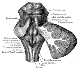
The rhomboid fossa is a rhombus-shaped depression that is the anterior part of the fourth ventricle. Its anterior wall, formed by the back of the pons and the medulla oblongata, constitutes the floor of the fourth ventricle.

The spinotectal tract arises in the spinothalamic tract and terminates in the inferior and superior colliculi.

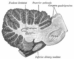
In the human brain, the superior cerebellar peduncle is a paired structure of white matter that connects the cerebellum to the midbrain. It consists mainly of efferent fibers, the cerebellothalamic tract that runs from a cerebellar hemisphere to the contralateral thalamus, and the cerebellorubral tract that runs from a cerebellar hemisphere to the red nucleus. It also contains afferent tracts, most prominent of which is the ventral spinocerebellar tract. Other afferent tracts are the trigeminothalamic fibers, tectocerebellar fibers, and noradrenergic fibers from the locus coeruleus. The superior peduncle emerges from the upper and medial parts of the white matter of each hemisphere and is placed under cover of the upper part of the cerebellum.

The middle cerebellar peduncle is a paired structure of the brain. It connects the pons to the cerebellum, with fibres originating from the pontine nucleus and travelling to the opposite hemisphere of the cerebellar cortex. It is supplied by the anterior inferior cerebellar artery (AICA) and branches from the basilar artery. It conveys information from the cerebrum and the pons to the cerebellum.

The superior medullary velum is a thin, transparent lamina of white matter which - together with the inferior medullary velum - forms the roof of the fourth ventricle. It extends between the two superior cerebellar peduncles. The lingula of cerebellum covers - and adheres to - its dorsal surface.

In the ventral cochlear nucleus (VCN), auditory nerve fibers enter the brain via the nerve root in the VCN. The ventral cochlear nucleus is divided into the anterior ventral (anteroventral) cochlear nucleus (AVCN) and the posterior ventral (posteroventral) cochlear nucleus (PVCN). In the VCN, auditory nerve fibers bifurcate, the ascending branch innervates the AVCN and the descending branch innervates the PVCN and then continue to the dorsal cochlear nucleus. The orderly innervation by auditory nerve fibers gives the AVCN a tonotopic organization along the dorsoventral axis. Fibers that carry information from the apex of the cochlea that are tuned to low frequencies contact neurons in the ventral part of the AVCN; those that carry information from the base of the cochlea that are tuned to high frequencies contact neurons in the dorsal part of the AVCN. Several populations of neurons populate the AVCN. Bushy cells receive input from auditory nerve fibers through particularly large endings called end bulbs of Held. They contact stellate cells through more conventional boutons.