Pretectal area | |
---|---|
Details | |
Part of | Midbrain |
Parts | anterior pretectal nucleus, medial pretectal nucleus, nucleus of the optic tract, olivary pretectal nucleus, posterior pretectal nucleus, posterior limitans, commissural pretectal area |
Identifiers | |
Latin | area praetectalis |
MeSH | D066250 |
NeuroNames | 467 |
NeuroLex ID | nlx_59721 |
TA98 | A14.1.08.505 A14.1.08.506 |
TA2 | 5739 |
FMA | 62402 |
Anatomical terms of neuroanatomy |
In neuroanatomy, the pretectal area, or pretectum, is a midbrain structure composed of seven nuclei and comprises part of the subcortical visual system. Through reciprocal bilateral projections from the retina, it is involved primarily in mediating behavioral responses to acute changes in ambient light such as the pupillary light reflex, the optokinetic reflex, and temporary changes to the circadian rhythm. [1] [2] [3] [4] [5] In addition to the pretectum's role in the visual system, the anterior pretectal nucleus has been found to mediate somatosensory and nociceptive information. [6] [7]
The pretectum is a bilateral group of highly interconnected nuclei located near the junction of the midbrain and forebrain. [8] The pretectum is generally classified as a midbrain structure, although because of its proximity to the forebrain it is sometimes classified as part of the caudal diencephalon (forebrain). [9] Within vertebrates, the pretectum is located directly anterior to the superior colliculus and posterior to the thalamus. It is situated above the periaqueductal grey and nucleus of the posterior commissure. [10]
Several nuclei have been identified within the pretectum, although their borders can be difficult to define and there has been debate over which regions should be included and their precise names. [1] [10] [11] The five primary nuclei are: the olivary pretectal nucleus (ON), the nucleus of the optic tract (NOT), and the anterior (NPA), medial (NPM), and posterior (NPP) pretectal nuclei. The NOT consists of relatively large cells and is located between the superior colliculi. The ON is located medial to the NOT and has a tail that extends between the NOT and NPP, which is ventral to the ON. [10] Two additional nuclei have also been identified: the posterior limitans (PLi) and the commisural pretectal area (CPA). [12] While these two regions have not been examined to the same extent as the five primary nuclei, research has shown both the PLi and CPA to receive retinal input, which suggests a role in processing visual information. [13]
The pretectum receives significant binocular input from photosensitive ganglion cells in the retina. In primates these afferents are bilateral [14] while in rodents they project from the contralateral retina. The majority of these retino-pretectal projections go to the ON and NOT [14] while other pretectal nuclei receive minor retinal input in mammals including the posterior, medial, and anterior pretectal nuclei. [1] [10] [15] [16]
The NOT receives input from several regions. From the thalamus the NOT receives inhibitory projections from GABA-producing neurons in the ipsilateral lateral geniculate nucleus and bilateral intergeniculate leaflets. The ipsilateral superficial suprachiasmatic nucleus and the medial, dorsal, and lateral terminal nuclei in the midbrain project onto the NOT. Fibers also project to the NOT from the nucleus prepositus hypoglossi in the medulla, the contralateral NOT, and from various cortical regions. [1] [17]
Many pretectal nuclei share targets of efferent projections. All pretectal nuclei, except for the ON, project to nuclei in the thalamus, subthalamus, superior colliculus, reticular formation, pons, and inferior olive. [10] Both the ON and the CPA have efferent projections to the Edinger-Westphal nucleus. The NPP and NPA both project to the pulvinar, the lateral posterior nucleus of the thalamus, and several precerebellar nuclei. [1]
The NOT has efferent projections to the zona incerta of the subthalamus, several nuclei of the pons, medulla, intralaminar nuclei, midbrain, and dorsal and ventral thalamic nuclei. Its bilateral inhibitory projections to the accessory optic system include connections to the lateral and medial terminal nuclei. Projections to the subthalamus are target toward the lateral geniculate nucleus and pulvinar. The NOT projects bilaterally to the superior colliculus, although the ipsilateral connections appear to be more dominant. In addition to these projections, the NOT projects to the vestibular and vestibulocerebellar relay nuclei. [1]
As part of the subcortical visual system, neurons within the pretectal nuclei respond to varying intensities of illuminance and are primarily involved in mediating non-conscious behavioral responses to acute changes in light. In general, these responses involve the initiation of optokinetic reflexes, although the pretectum can also regulate nociception and REM sleep. [12]
The pupillary light reflex is mediated by the pretectum. [2] This reflex is responsible for the constriction of the pupils upon light's entering the eye. Several pretectal nuclei, in particular the ON, receive illuminance information from the ipsilateral side of the retinas of both eyes via the optic tract. Nuclei in the ON are known to gradually increase in activation in response to increasing levels of illuminance. This information is then relayed directly to the Edinger-Westphal nucleus, which proceeds to relay the command to constrict the pupils to the pupillary sphincter via the ciliary ganglion. [4] [18]
Pretectal nuclei, in particular the NOT, are involved in coordinating eye movements during smooth pursuit. These movements allow the eye to closely follow a moving object and to catch up to an object after an unexpected change in direction or velocity. Direction-sensitive retinal slip neurons within the NOT provide ipsiversive horizontal retinal error information to the cortex through the inferior olive. During the day, this information is sensed and relayed by neurons with large receptive fields, whereas parafoveal neurons with small receptive fields do so in the dark. It is through this pathway that the NOT is able to provide retinal error information to guide eye movements. [1] [17] [19] In addition to its role in maintaining smooth pursuit, the pretectum is activated during the optokinetic nystagmus in which the eye returns to a central, forward-facing position after an object it was following passes out of the field of vision. [20]
Part of the pretectum, particularly the NOT and NPP, are implicated in the accommodation reflex by which the eye maintains focus. [21] Proprioceptive information from the retina reaches the pretectum via the occulomotor nerve and the trigeminal nerve. From that point, the mechanism by which the eye maintains focus through muscular contractions of the retina is similar to that of the pupillary light reflex. [4]
The NPA participates in the active diminishing of the perception of pain stimuli (antinociception). [7] Although the mechanism by which the NPA alters an organism's response to painful stimuli is not fully known, research has shown that activity in the ventral NPA triggers cholinergic and serotonergic neurons. These neurons activate descending pathways that synapse in the spinal cord and inhibit nociceptive cells in the dorsal horn. [22] In addition to its direct antionociceptive mechanism, the NPA projects onto brain regions that, through connections to the somatosensory cortex, regulate the perception of painful stimuli. Two of these regions that the NPA is known to project to are the zona incerta and posterior thalamic nucleus. Regions of the NPA may be specialized to respond to different types of pain. Research has found that the dorsal NPA best diminished the perception of brief pain whereas the ventral NPA reduced the perception of chronic pain. [23] Because of its role in the reduction of chronic pain, abnormal activity of the NPA is thought to be implicated in central pain syndrome. [24]
Multiple pretectal nuclei may be involved in regulating REM sleep and sleep behaviors. Research has shown that the pretectum, in conjunction with the superior colliculus, may be responsible for causing non-circadian changes in REM sleep behaviors. [25] Pretectal nuclei receiving retinal input, in particular the NOT and the NPP, have been shown to be partially responsible for initiating REM sleep in albino rats. [5] The discovery of projections from the pretectum to several thalamic nuclei involved in cortical activation during REM sleep, to be specific the projection to the superchiasmatic nucleus, which is part of a known REM sleep regulatory mechanism, supports this hypothesis. [12]
The thalamus is a large mass of gray matter on the lateral walls of the third ventricle forming the dorsal part of the diencephalon. Nerve fibers project out of the thalamus to the cerebral cortex in all directions, known as the thalamocortical radiations, allowing hub-like exchanges of information. It has several functions, such as the relaying of sensory and motor signals to the cerebral cortex and the regulation of consciousness, sleep, and alertness.
Articles related to anatomy include:
The brainstem is the stalk-like part of the brain that interconnects the cerebrum and diencephalon with the spinal cord. In the human brain, the brainstem is composed of the midbrain, the pons, and the medulla oblongata. The midbrain is continuous with the thalamus of the diencephalon through the tentorial notch.
The visual system is the physiological basis of visual perception. The system detects, transduces and interprets information concerning light within the visible range to construct an image and build a mental model of the surrounding environment. The visual system is associated with the eye and functionally divided into the optical system and the neural system.
In neuroanatomy, the lateral geniculate nucleus is a structure in the thalamus and a key component of the mammalian visual pathway. It is a small, ovoid, ventral projection of the thalamus where the thalamus connects with the optic nerve. There are two LGNs, one on the left and another on the right side of the thalamus. In humans, both LGNs have six layers of neurons alternating with optic fibers.
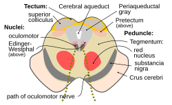
The midbrain or mesencephalon is the rostral-most portion of the brainstem connecting the diencephalon and cerebrum with the pons. It consists of the cerebral peduncles, tegmentum, and tectum.
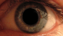
The pupillary light reflex (PLR) or photopupillary reflex is a reflex that controls the diameter of the pupil, in response to the intensity (luminance) of light that falls on the retinal ganglion cells of the retina in the back of the eye, thereby assisting in adaptation of vision to various levels of lightness/darkness. A greater intensity of light causes the pupil to constrict, whereas a lower intensity of light causes the pupil to dilate. Thus, the pupillary light reflex regulates the intensity of light entering the eye. Light shone into one eye will cause both pupils to constrict.
The fibers of the oculomotor nerve arise from a nucleus in the midbrain, which lies in the gray substance of the floor of the cerebral aqueduct and extends in front of the aqueduct for a short distance into the floor of the third ventricle. From this nucleus the fibers pass forward through the tegmentum, the red nucleus, and the medial part of the substantia nigra, forming a series of curves with a lateral convexity, and emerge from the oculomotor sulcus on the medial side of the cerebral peduncle.
In neuroanatomy, the superior colliculus is a structure lying on the roof of the mammalian midbrain. In non-mammalian vertebrates, the homologous structure is known as the optic tectum or optic lobe. The adjective form tectal is commonly used for both structures.
The inferior colliculus (IC) is the principal midbrain nucleus of the auditory pathway and receives input from several peripheral brainstem nuclei in the auditory pathway, as well as inputs from the auditory cortex. The inferior colliculus has three subdivisions: the central nucleus, a dorsal cortex by which it is surrounded, and an external cortex which is located laterally. Its bimodal neurons are implicated in auditory-somatosensory interaction, receiving projections from somatosensory nuclei. This multisensory integration may underlie a filtering of self-effected sounds from vocalization, chewing, or respiration activities.
The reticular formation is a set of interconnected nuclei that are located in the brainstem, hypothalamus, and other regions. It is not anatomically well defined, because it includes neurons located in different parts of the brain. The neurons of the reticular formation make up a complex set of networks in the core of the brainstem that extend from the upper part of the midbrain to the lower part of the medulla oblongata. The reticular formation includes ascending pathways to the cortex in the ascending reticular activating system (ARAS) and descending pathways to the spinal cord via the reticulospinal tracts.
The Edinger–Westphal nucleus is one of two nuclei of the oculomotor nerve and is located in the midbrain. It receives afferents from both pretectal nuclei. It contains parasympathetic pre-ganglionic neuron cell bodies that synapse in the ciliary ganglion. It contributes the autonomic, parasympathetic component to the oculomotor nerve, ultimately providing innervation to the iris sphincter muscle and ciliary muscle to mediate the pupillary light reflex and accommodation, respectively.
Intrinsically photosensitive retinal ganglion cells (ipRGCs), also called photosensitive retinal ganglion cells (pRGC), or melanopsin-containing retinal ganglion cells (mRGCs), are a type of neuron in the retina of the mammalian eye. The presence of an additional photoreceptor was first suspected in 1927 when mice lacking rods and cones still responded to changing light levels through pupil constriction; this suggested that rods and cones are not the only light-sensitive tissue. However, it was unclear whether this light sensitivity arose from an additional retinal photoreceptor or elsewhere in the body. Recent research has shown that these retinal ganglion cells, unlike other retinal ganglion cells, are intrinsically photosensitive due to the presence of melanopsin, a light-sensitive protein. Therefore, they constitute a third class of photoreceptors, in addition to rod and cone cells.
The zona incerta (ZI) is a horizontally elongated region of gray matter in the subthalamus below the thalamus. Its connections project extensively over the brain from the cerebral cortex down into the spinal cord.
The optokinetic reflex (OKR), also referred to as the optokinetic response, or optokinetic nystagmus (OKN), is a compensatory reflex that supports visual image stabilization. The purpose of OKR is to prevent image blur on the retina that would otherwise occur when an animal moves its head or navigates through its environment. This is achieved by the reflexive movement of the eyes in the same direction as image motion, so as to minimize the relative motion of the visual scene on the eye. OKR is best evoked by slow, rotational motion, and operates in coordination with several complementary reflexes that also support image stabilization, including the vestibulo-ocular reflex (VOR).
The medial dorsal nucleus is a large nucleus in the thalamus.
The isothalamus is a division used by some researchers in describing the thalamus.
The parabrachial nuclei, also known as the parabrachial complex, are a group of nuclei in the dorsolateral pons that surrounds the superior cerebellar peduncle as it enters the brainstem from the cerebellum. They are named from the Latin term for the superior cerebellar peduncle, the brachium conjunctivum. In the human brain, the expansion of the superior cerebellar peduncle expands the parabrachial nuclei, which form a thin strip of grey matter over most of the peduncle. The parabrachial nuclei are typically divided along the lines suggested by Baxter and Olszewski in humans, into a medial parabrachial nucleus and lateral parabrachial nucleus. These have in turn been subdivided into a dozen subnuclei: the superior, dorsal, ventral, internal, external and extreme lateral subnuclei; the lateral crescent and subparabrachial nucleus along the ventrolateral margin of the lateral parabrachial complex; and the medial and external medial subnuclei
Lateral pulvinar nucleus is one of four traditionally anatomically distinguished nuclei of the pulvinar of the thalamus. The other three nuclei of the pulvinar are called anterior, inferior and medial pulvinar nuclei.
Medial pulvinar nucleus is one of four traditionally anatomically distinguished nuclei of the pulvinar of the thalamus. The other three nuclei of the pulvinar are called lateral, inferior and anterior pulvinar nuclei.