Grid illusion

Last updated
An example of the Hermann grid illusion. Dark blobs appear at the intersections HermannGrid.svg
An example of the Hermann grid illusion. Dark blobs appear at the intersections

A grid illusion is any kind of grid that deceives a person's vision. The two most common types of grid illusions are the Hermann grid illusion and the scintillating grid illusion.

Contents

Hermann grid illusion

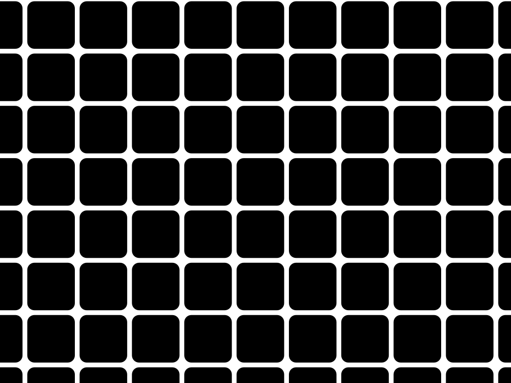
The Hermann grid illusion is an optical illusion reported by Ludimar Hermann in 1870. [1] The illusion is characterized by "ghostlike" grey blobs perceived at the intersections of a white (or light-colored) grid on a black background. The grey blobs disappear when looking directly at an intersection.

Scintillating grid illusion

An example of the scintillating grid illusion. Dark dots seem to appear and disappear at intersections. Grid illusion.svg
An example of the scintillating grid illusion. Dark dots seem to appear and disappear at intersections.

The scintillating grid illusion is an optical illusion, discovered by E. and B. Lingelbach and M. Schrauf in 1994. [2] It is often considered a variation of the Hermann grid illusion but possesses different properties. [2] [3]

It is constructed by superimposing white discs on the intersections of orthogonal gray bars on a black background. Dark dots seem to appear and disappear rapidly at random intersections, hence the label "scintillating". When a person keeps their eyes directly on a single intersection, the dark dot does not appear. The dark dots disappear if one is too close to or too far from the image.

Differences between the scintillating and Hermann grid illusions

The difference between the Scintillating Grid Illusion and the Hermann Grid Illusion is that the former has dots already in place at the intersections, which is not the case for the latter. Since, at first sight, the graphs appear similar, the two illusions are occasionally confused. But the scintillating illusion does not occur with an isolated intersection, as is the case for the Hermann grid; observations suggest that a minimum of 3 × 3 evenly spaced intersections with superimposed discs are required to produce the effect. This requirement suggests the participation of global processes of the kind proposed for the linking and grouping of features in an image, in addition to local processes. [4]

Theories

The effect of both optical illusions is often explained by a neural process called lateral inhibition. [5] The intensity at a point in the visual system is not simply the result of a single receptor, but the result of a group of receptors which respond to the presentation of stimuli in what is called a receptive field.

A retinal ganglion cell pools the inputs of several photoreceptors over an area of the retina; the area in physical space to which the photoreceptors respond is the ganglion cell's "receptive field". In the center of a so-called on-center receptive field, the individual photoreceptors excite the ganglion cell when they detect increased luminance; the photoreceptors in the surrounding area inhibit the ganglion cell. Thus, since a point at an intersection is surrounded by more areas of intensity than a point at the middle of a line, the intersection appears darker due to the increased inhibition.

There is strong evidence that the retinal ganglion cell theory is untenable. For example, making the lines of the grid wavy rather than straight eliminates both the Hermann grid and scintillating grid illusions. [6] [7] [8] [9] [10] [11] The Baumgartner / RGC theory does not predict this outcome. Lateral inhibition theory also can not account for the fact that the Hermann grid illusion is perceived over a range of bar widths. [8] Lateral inhibition theory would predict that decreasing the size of the grid (and therefore decreasing the amount of inhibition at the intersection) would eradicate the illusory effect. One alternative explanation is that the illusion is due to S1 type simple cells in the visual cortex. [8]

See also

Related Research Articles

<span class="mw-page-title-main">Retina</span> Part of the eye

The retina is the innermost, light-sensitive layer of tissue of the eye of most vertebrates and some molluscs. The optics of the eye create a focused two-dimensional image of the visual world on the retina, which then processes that image within the retina and sends nerve impulses along the optic nerve to the visual cortex to create visual perception. The retina serves a function which is in many ways analogous to that of the film or image sensor in a camera.

<span class="mw-page-title-main">Optical illusion</span> Visually perceived images that differ from objective reality

In visual perception, an optical illusion is an illusion caused by the visual system and characterized by a visual percept that arguably appears to differ from reality. Illusions come in a wide variety; their categorization is difficult because the underlying cause is often not clear but a classification proposed by Richard Gregory is useful as an orientation. According to that, there are three main classes: physical, physiological, and cognitive illusions, and in each class there are four kinds: Ambiguities, distortions, paradoxes, and fictions. A classical example for a physical distortion would be the apparent bending of a stick half immerged in water; an example for a physiological paradox is the motion aftereffect. An example for a physiological fiction is an afterimage. Three typical cognitive distortions are the Ponzo, Poggendorff, and Müller-Lyer illusion. Physical illusions are caused by the physical environment, e.g. by the optical properties of water. Physiological illusions arise in the eye or the visual pathway, e.g. from the effects of excessive stimulation of a specific receptor type. Cognitive visual illusions are the result of unconscious inferences and are perhaps those most widely known.

<span class="mw-page-title-main">Visual system</span> Body parts responsible for sight

The visual system comprises the sensory organ and parts of the central nervous system which gives organisms the sense of sight as well as enabling the formation of several non-image photo response functions. It detects and interprets information from the optical spectrum perceptible to that species to "build a representation" of the surrounding environment. The visual system carries out a number of complex tasks, including the reception of light and the formation of monocular neural representations, colour vision, the neural mechanisms underlying stereopsis and assessment of distances to and between objects, the identification of a particular object of interest, motion perception, the analysis and integration of visual information, pattern recognition, accurate motor coordination under visual guidance, and more. The neuropsychological side of visual information processing is known as visual perception, an abnormality of which is called visual impairment, and a complete absence of which is called blindness. Non-image forming visual functions, independent of visual perception, include the pupillary light reflex and circadian photoentrainment.

<span class="mw-page-title-main">Sensory nervous system</span> Part of the nervous system responsible for processing sensory information

The sensory nervous system is a part of the nervous system responsible for processing sensory information. A sensory system consists of sensory neurons, neural pathways, and parts of the brain involved in sensory perception and interoception. Commonly recognized sensory systems are those for vision, hearing, touch, taste, smell, balance and visceral sensation. Sense organs are transducers that convert data from the outer physical world to the realm of the mind where people interpret the information, creating their perception of the world around them.

<span class="mw-page-title-main">Lateral geniculate nucleus</span> Component of the visual system in the brains thalamus

In neuroanatomy, the lateral geniculate nucleus is a structure in the thalamus and a key component of the mammalian visual pathway. It is a small, ovoid, ventral projection of the thalamus where the thalamus connects with the optic nerve. There are two LGNs, one on the left and another on the right side of the thalamus. In humans, both LGNs have six layers of neurons alternating with optic fibers.

<span class="mw-page-title-main">Photoreceptor cell</span> Type of neuroepithelial cell

A photoreceptor cell is a specialized type of neuroepithelial cell found in the retina that is capable of visual phototransduction. The great biological importance of photoreceptors is that they convert light into signals that can stimulate biological processes. To be more specific, photoreceptor proteins in the cell absorb photons, triggering a change in the cell's membrane potential.

<span class="mw-page-title-main">Retinal ganglion cell</span> Type of cell within the eye

A retinal ganglion cell (RGC) is a type of neuron located near the inner surface of the retina of the eye. It receives visual information from photoreceptors via two intermediate neuron types: bipolar cells and retina amacrine cells. Retina amacrine cells, particularly narrow field cells, are important for creating functional subunits within the ganglion cell layer and making it so that ganglion cells can observe a small dot moving a small distance. Retinal ganglion cells collectively transmit image-forming and non-image forming visual information from the retina in the form of action potential to several regions in the thalamus, hypothalamus, and mesencephalon, or midbrain.

<span class="mw-page-title-main">Sensory neuron</span> Nerve cell that converts environmental stimuli into corresponding internal stimuli

Sensory neurons, also known as afferent neurons, are neurons in the nervous system, that convert a specific type of stimulus, via their receptors, into action potentials or graded receptor potentials. This process is called sensory transduction. The cell bodies of the sensory neurons are located in the dorsal ganglia of the spinal cord.

The receptive field, or sensory space, is a delimited medium where some physiological stimuli can evoke a sensory neuronal response in specific organisms.

<span class="mw-page-title-main">Retina bipolar cell</span> Type of neuron

As a part of the retina, bipolar cells exist between photoreceptors and ganglion cells. They act, directly or indirectly, to transmit signals from the photoreceptors to the ganglion cells.

<span class="mw-page-title-main">Melanopsin</span> Mammalian protein found in Homo sapiens

Melanopsin is a type of photopigment belonging to a larger family of light-sensitive retinal proteins called opsins and encoded by the gene Opn4. In the mammalian retina, there are two additional categories of opsins, both involved in the formation of visual images: rhodopsin and photopsin in the rod and cone photoreceptor cells, respectively.

<span class="mw-page-title-main">Motion perception</span> Inferring the speed and direction of objects

Motion perception is the process of inferring the speed and direction of elements in a scene based on visual, vestibular and proprioceptive inputs. Although this process appears straightforward to most observers, it has proven to be a difficult problem from a computational perspective, and difficult to explain in terms of neural processing.

<span class="mw-page-title-main">Amacrine cell</span> Interneuron cells in the retina of the eye

In the anatomy of the eye, amacrine cells are interneurons in the retina. They are named from Greek a– 'non', makr– 'long', and in– 'fiber', because of their short neuronal processes. Amacrine cells are inhibitory neurons, and they project their dendritic arbors onto the inner plexiform layer (IPL), they interact with retinal ganglion cells, and bipolar cells or both of these.

<span class="mw-page-title-main">Retina horizontal cell</span>

Horizontal cells are the laterally interconnecting neurons having cell bodies in the inner nuclear layer of the retina of vertebrate eyes. They help integrate and regulate the input from multiple photoreceptor cells. Among their functions, horizontal cells are believed to be responsible for increasing contrast via lateral inhibition and adapting both to bright and dim light conditions. Horizontal cells provide inhibitory feedback to rod and cone photoreceptors. They are thought to be important for the antagonistic center-surround property of the receptive fields of many types of retinal ganglion cells.

<span class="mw-page-title-main">Hering illusion</span> Geometrical-optical illusion

The Hering illusion is one of the geometrical-optical illusions and was discovered by the German physiologist Ewald Hering in 1861. When two straight and parallel lines are presented in front of radial background, the lines appear as if they were bowed outwards. The Orbison illusion is one of its variants, while the Wundt illusion produces a similar, but inverted effect.

Intrinsically photosensitive retinal ganglion cells (ipRGCs), also called photosensitive retinal ganglion cells (pRGC), or melanopsin-containing retinal ganglion cells (mRGCs), are a type of neuron in the retina of the mammalian eye. The presence of ipRGCs was first suspected in 1927 when rodless, coneless mice still responded to a light stimulus through pupil constriction, This implied that rods and cones are not the only light-sensitive neurons in the retina. Yet research on these cells did not advance until the 1980s. Recent research has shown that these retinal ganglion cells, unlike other retinal ganglion cells, are intrinsically photosensitive due to the presence of melanopsin, a light-sensitive protein. Therefore, they constitute a third class of photoreceptors, in addition to rod and cone cells.

<span class="mw-page-title-main">Lateral inhibition</span> Capacity of an excited neuron to reduce activity of its neighbors

In neurobiology, lateral inhibition is the capacity of an excited neuron to reduce the activity of its neighbors. Lateral inhibition disables the spreading of action potentials from excited neurons to neighboring neurons in the lateral direction. This creates a contrast in stimulation that allows increased sensory perception. It is also referred to as lateral antagonism and occurs primarily in visual processes, but also in tactile, auditory, and even olfactory processing. Cells that utilize lateral inhibition appear primarily in the cerebral cortex and thalamus and make up lateral inhibitory networks (LINs). Artificial lateral inhibition has been incorporated into artificial sensory systems, such as vision chips, hearing systems, and optical mice. An often under-appreciated point is that although lateral inhibition is visualised in a spatial sense, it is also thought to exist in what is known as "lateral inhibition across abstract dimensions." This refers to lateral inhibition between neurons that are not adjacent in a spatial sense, but in terms of modality of stimulus. This phenomenon is thought to aid in colour discrimination.

Feature detection is a process by which the nervous system sorts or filters complex natural stimuli in order to extract behaviorally relevant cues that have a high probability of being associated with important objects or organisms in their environment, as opposed to irrelevant background or noise.

<span class="mw-page-title-main">Parasol cell</span>

A parasol cell, sometimes called an M cell or M ganglion cell, is one type of retinal ganglion cell (RGC) located in the ganglion cell layer of the retina. These cells project to magnocellular cells in the lateral geniculate nucleus (LGN) as part of the magnocellular pathway in the visual system. They have large cell bodies as well as extensive branching dendrite networks and as such have large receptive fields. Relative to other RGCs, they have fast conduction velocities. While they do show clear center-surround antagonism, they receive no information about color. Parasol ganglion cells contribute information about the motion and depth of objects to the visual system.

<i>Pflügers Archiv: European Journal of Physiology</i> Academic journal

Pflügers Archiv: European Journal of Physiology is a peer-reviewed scientific journal in the field of physiology. A continuation of a journal founded in 1868 by the German physiologist, Eduard Friedrich Wilhelm Pflüger, Pflügers Archiv is the oldest physiological journal. Pflügers Archiv is currently published by Springer, with 11 issues per year.

References

  1. Hermann L (1870). "Eine Erscheinung simultanen Contrastes". Pflügers Archiv für die gesamte Physiologie. 3: 13–15. doi:10.1007/BF01855743. S2CID   41109941.
  2. 1 2 Schrauf, M.; Lingelbach, B.; Lingelbach, E.; Wist, E. R. (1995). "The Hermann Grid and the scintillation effect". Perception. 24 Suppl. A: 88–89.
  3. Schrauf, M.; Lingelbach, B.; Wist, E. R. (1997). "The Scintillating Grid Illusion". Vision Research. 37 (8): 1033–1038. doi: 10.1016/S0042-6989(96)00255-6 . PMID   9196721.
  4. Alexander, D.M.; Van Leeuwen, C. (2010). "Mapping of contextual modulation in the population response of primary visual cortex". Cognitive Neurodynamics. 4 (1): 1–24. doi:10.1007/s11571-009-9098-9. PMC   2837531 . PMID   19898958.
  5. Baumgartner G (1960). "Indirekte Größenbestimmung der rezeptiven Felder der Retina beim Menschen mittels der Hermannschen Gittertäuschung". Pflügers Archiv für die gesamte Physiologie. 272: 21–22. doi:10.1007/BF00680926. S2CID   45209673.
  6. Lingelbach B, Block B, Hatzky B, Reisinger E (1985). "The Hermann grid illusion–retinal or cortical?". Perception. 14 (1): A7.
  7. Geier J, Bernáth L (2004). "Stopping the Hermann grid illusion by simple sine distortion". Perception. Malden Ma: Blackwell. pp. 33–53. ISBN   0631224211.
  8. 1 2 3 Schiller, Peter H.; Carvey, Christina E. (2005). "The Hermann grid illusion revisited". Perception. 34 (11): 1375–1397. doi:10.1068/p5447. PMID   16355743. S2CID   15740144. Archived from the original on 2011-12-12. Retrieved 2011-10-03.
  9. Geier J, Bernáth L, Hudák M, Séra L (2008). "Straightness as the main factor of the Hermann grid illusion". Perception. 37 (5): 651–665. doi:10.1068/p5622. PMID   18605141. S2CID   21028439.
  10. Geier, János (2008). "Stopping the Hermann grid illusion by sine distortion".
  11. Bach, Michael (2008). "Die Hermann-Gitter-Täuschung: Lehrbucherklärung widerlegt (The Hermann grid illusion: the classic textbook interpretation is obsolete)". Ophthalmologe. 106 (10): 913–917. doi:10.1007/s00347-008-1845-5. PMID   18830602.