Medial dorsal nucleus

Last updated
Medial dorsal nucleus
ThalamicNuclei.svg
Thalmus.png
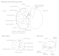
Thalamic nuclei (right thalamus from above)
Details
Identifiers
Latin nucleus mediodorsalis thalami
MeSH D020645
NeuroNames 312
NeuroLex ID birnlex_1543
TA98 A14.1.08.622
TA2 5681
FMA 62156
Anatomical terms of neuroanatomy

The medial dorsal nucleus (or mediodorsal nucleus of thalamus, or dorsal medial nucleus [1] ) is a large nucleus in the thalamus. [2] It is separated from the other thalamic nuclei by the internal medullary lamina.

Contents

The medial dorsal nucleus is interconnected with the prefrontal cortex, therefore involved in prefrontal functions. Damage to the interconnected tract or the nucleus itself will result in similar damage to the prefrontal cortex. [1] It is also believed to play a role in memory. [3]

Structure

It relays inputs from the amygdala and olfactory cortex and projects to the prefrontal cortex and the limbic system and in turn relays them to the prefrontal association cortex. As a result, it plays a crucial role in attention, planning, organization, abstract thinking, multi-tasking, and active memory.[ citation needed ]

The connections of the medial dorsal nucleus have even been used to delineate the prefrontal cortex of the Göttingen minipig brain. [4]

By stereology the number of brain cells in the region has been estimated at around 6.43 million neurons in the adult human brain and 36.3 million glial cells, with the newborn having quite different numbers: around 11.2 million neurons and 10.6 million glial cells. [5]

Function

Pain processing

While both the ventral and medial dorsal nuclei process pain, the medial dorsal nucleus bypasses primary cortices, sending their axons directly to secondary and association cortices. The cells also send axons directly to many parts of the brain, including nuclei of the limbic system such as the lateral nucleus of the amygdala, the anterior cingulate, and the hippocampus. This part of the sensory system, known as the non-classical or extralemniscal system is less accurate, and less detailed in regards to sensory signal analysis. This processing is known colloquially as "fast and dirty" rather than the "slow and accurate" system of classical or lemniscal system. This pathway activates parts of the brain that evoke emotional responses.[ citation needed ]

Saccadic efference copy

This nucleus is also presumed to play a role in monitoring internal movements of the eye. Specifically, its function is to relay the information about how the eyes will be moved (efference copy, also known as corollary discharge) from the superior colliculus to the frontal eye fields (FEF) in order to aid the neurons in FEF to change their receptive fields to where the visual stimuli will appear after the saccade. [6]

Clinical significance

Damage to the medial dorsal nucleus has been associated with Korsakoff's syndrome. [7]

Additional images

Related Research Articles

<span class="mw-page-title-main">Thalamus</span> Structure within the brain

The thalamus is a large mass of gray matter on the lateral walls of the third ventricle forming the dorsal part of the diencephalon. Nerve fibers project out of the thalamus to the cerebral cortex in all directions, known as the thalamocortical radiations, allowing hub-like exchanges of information. It has several functions, such as the relaying of sensory and motor signals to the cerebral cortex and the regulation of consciousness, sleep, and alertness.

<span class="mw-page-title-main">Lateral geniculate nucleus</span> Component of the visual system in the brains thalamus

In neuroanatomy, the lateral geniculate nucleus is a structure in the thalamus and a key component of the mammalian visual pathway. It is a small, ovoid, ventral projection of the thalamus where the thalamus connects with the optic nerve. There are two LGNs, one on the left and another on the right side of the thalamus. In humans, both LGNs have six layers of neurons alternating with optic fibers.

<span class="mw-page-title-main">Cingulate cortex</span> Part of the brain within the cerebral cortex

The cingulate cortex is a part of the brain situated in the medial aspect of the cerebral cortex. The cingulate cortex includes the entire cingulate gyrus, which lies immediately above the corpus callosum, and the continuation of this in the cingulate sulcus. The cingulate cortex is usually considered part of the limbic lobe.

<span class="mw-page-title-main">Dorsal column–medial lemniscus pathway</span> Sensory spinal pathway

The dorsal column–medial lemniscus pathway (DCML) (also known as the posterior column-medial lemniscus pathway is the major sensory pathway of the central nervous system that conveys sensations of fine touch, vibration, two-point discrimination, and proprioception from the skin and joints. It transmits this information to the somatosensory cortex of the postcentral gyrus in the parietal lobe of the brain. The pathway receives information from sensory receptors throughout the body, and carries this in the gracile fasciculus and the cuneate fasciculus, tracts that make up the white matter dorsal columns of the spinal cord. At the level of the medulla oblongata, the fibers of the tracts decussate and are continued in the medial lemniscus, on to the thalamus and relayed from there through the internal capsule and transmitted to the somatosensory cortex. The name dorsal-column medial lemniscus comes from the two structures that carry the sensory information: the dorsal columns of the spinal cord, and the medial lemniscus in the brainstem.

<span class="mw-page-title-main">Pulvinar nuclei</span>

The pulvinar nuclei or nuclei of the pulvinar are the nuclei located in the thalamus. As a group they make up the collection called the pulvinar of the thalamus, usually just called the pulvinar.

<span class="mw-page-title-main">Reticular formation</span> Spinal trigeminal nucleus

The reticular formation is a set of interconnected nuclei that is located in the brainstem, hypothalamus, and other regions. It is not anatomically well defined, because it includes neurons located in different parts of the brain. The neurons of the reticular formation make up a complex set of networks in the core of the brainstem that extend from the upper part of the midbrain to the lower part of the medulla oblongata.

<span class="mw-page-title-main">Thalamocortical radiations</span> Neural pathways between the thalamus and cerebral cortex

In neuroanatomy, thalamocortical radiations, also known as thalamocortical fibers, are the efferent fibers that project from the thalamus to distinct areas of the cerebral cortex. They form fiber bundles that emerge from the lateral surface of the thalamus.

<span class="mw-page-title-main">Dentate nucleus</span> Nucleus in the centre of each cerebellar hemisphere

The dentate nucleus is a cluster of neurons, or nerve cells, in the central nervous system that has a dentate – tooth-like or serrated – edge. It is located within the deep white matter of each cerebellar hemisphere, and it is the largest single structure linking the cerebellum to the rest of the brain. It is the largest and most lateral, or farthest from the midline, of the four pairs of deep cerebellar nuclei, the others being the globose and emboliform nuclei, which together are referred to as the interposed nucleus, and the fastigial nucleus. The dentate nucleus is responsible for the planning, initiation and control of voluntary movements. The dorsal region of the dentate nucleus contains output channels involved in motor function, which is the movement of skeletal muscle, while the ventral region contains output channels involved in nonmotor function, such as conscious thought and visuospatial function.

<span class="mw-page-title-main">Medial geniculate nucleus</span>

The medial geniculate nucleus (MGN) or medial geniculate body (MGB) is part of the auditory thalamus and represents the thalamic relay between the inferior colliculus (IC) and the auditory cortex (AC). It is made up of a number of sub-nuclei that are distinguished by their neuronal morphology and density, by their afferent and efferent connections, and by the coding properties of their neurons. It is thought that the MGN influences the direction and maintenance of attention.

<span class="mw-page-title-main">Septal area</span> Area in the lower, posterior part of the medial surface of the frontal lobe

The septal area, consisting of the lateral septum and medial septum, is an area in the lower, posterior part of the medial surface of the frontal lobe, and refers to the nearby septum pellucidum.

The zona incerta (ZI) is a horizontally elongated small nucleus that separates the larger subthalamic nucleus from the thalamus. Its connections project extensively over the brain from the cerebral cortex down into the spinal cord.

<span class="mw-page-title-main">Primate basal ganglia</span>

The basal ganglia form a major brain system in all vertebrates, but in primates there are special differentiating features. The basal ganglia include the striatum, pallidus, substantia nigra and subthalamic nucleus. In primates the pallidus is divided into an external and internal globus pallidus, the external globus pallidus is present in other mammals but not the internal globus pallidus. Also in primates, the dorsal striatum is divided by a large nerve tract called the internal capsule into two masses named the caudate nucleus and the putamen. These differences contribute to a complex circuitry of connections between the striatum and cortex that is specific to primates, reflecting different functions in primate cortical areas.

The amygdalofugal pathway is one of the three major efferent pathways of the amygdala, meaning that it is one of the three principal pathways by which fibers leave the amygdala. It leads from the basolateral nucleus and central nucleus of the amygdala. The amygdala is a limbic structure in the medial temporal lobe of the brain. The other main efferent pathways from the amygdala are the stria terminalis and anterior commissure.

The mammillothalamic tract is an efferent pathway of the mammillary body which projects to the anterior nuclei of the thalamus. It consists of heavily myelinated fibres. It is part of a brain circuit involved in spatial memory.

The isothalamus is a division used by some researchers in describing the thalamus.

<span class="mw-page-title-main">Synaptic gating</span>

Synaptic gating is the ability of neural circuits to gate inputs by either suppressing or facilitating specific synaptic activity. Selective inhibition of certain synapses has been studied thoroughly, and recent studies have supported the existence of permissively gated synaptic transmission. In general, synaptic gating involves a mechanism of central control over neuronal output. It includes a sort of gatekeeper neuron, which has the ability to influence transmission of information to selected targets independently of the parts of the synapse upon which it exerts its action.

Recurrent thalamo-cortical resonance or Thalamocortical oscillation is an observed phenomenon of oscillatory neural activity between the thalamus and various cortical regions of the brain. It is proposed by Rodolfo Llinas and others as a theory for the integration of sensory information into the whole of perception in the brain. Thalamocortical oscillation is proposed to be a mechanism of synchronization between different cortical regions of the brain, a process known as temporal binding. This is possible through the existence of thalamocortical networks, groupings of thalamic and cortical cells that exhibit oscillatory properties.

The trisynaptic circuit or trisynaptic loop is a relay of synaptic transmission in the hippocampus. The trisynaptic circuit is a neural circuit in the hippocampus, which is made up of three major cell groups: granule cells in the dentate gyrus, pyramidal neurons in CA3, and pyramidal neurons in CA1. The hippocampal relay involves 3 main regions within the hippocampus which are classified according to their cell type and projection fibers. The first projection of the hippocampus occurs between the entorhinal cortex (EC) and the dentate gyrus (DG). The entorhinal cortex transmits its signals from the parahippocampal gyrus to the dentate gyrus via granule cell fibers known collectively as the perforant path. The dentate gyrus then synapses on pyramidal cells in CA3 via mossy cell fibers. CA3 then fires to CA1 via Schaffer collaterals which synapse in the subiculum and are carried out through the fornix. Collectively the dentate gyrus, CA1 and CA3 of the hippocampus compose the trisynaptic loop.

<span class="mw-page-title-main">Parabrachial nuclei</span>

The parabrachial nuclei, also known as the parabrachial complex, are a group of nuclei in the dorsolateral pons that surrounds the superior cerebellar peduncle as it enters the brainstem from the cerebellum. They are named from the Latin term for the superior cerebellar peduncle, the brachium conjunctivum. In the human brain, the expansion of the superior cerebellar peduncle expands the parabrachial nuclei, which form a thin strip of grey matter over most of the peduncle. The parabrachial nuclei are typically divided along the lines suggested by Baxter and Olszewski in humans, into a medial parabrachial nucleus and lateral parabrachial nucleus. These have in turn been subdivided into a dozen subnuclei: the superior, dorsal, ventral, internal, external and extreme lateral subnuclei; the lateral crescent and subparabrachial nucleus along the ventrolateral margin of the lateral parabrachial complex; and the medial and external medial subnuclei

References

  1. 1 2 Vanderah, Todd W.; Gould, Douglas J.; Nolte, John (2016). Nolte's The human brain: an introduction to its functional anatomy (7th ed.). Philadelphia, PA: Elsevier. pp. 408–409. ISBN   978-1-4557-2859-6.
  2. Mitchell AS, Chakraborty S (2013). "What does the mediodorsal thalamus do?". Front Syst Neurosci. 7: 37. doi: 10.3389/fnsys.2013.00037 . PMC   3738868 . PMID   23950738.
  3. Li XB, Inoue T, Nakagawa S, Koyama T (May 2004). "Effect of mediodorsal thalamic nucleus lesion on contextual fear conditioning in rats". Brain Res. 1008 (2): 261–72. doi:10.1016/j.brainres.2004.02.038. PMID   15145764. S2CID   36284389.
  4. Jacob Jelsing; Anders Hay-Schmidt; Tim Dyrby; Ralf Hemmingsen; Harry B. M. Uylings; Bente Pakkenberg (2006). "The prefrontal cortex in the Göttingen minipig brain defined by neural projection criteria and cytoarchitecture". Brain Research Bulletin . 70 (4–6): 322–336. doi:10.1016/j.brainresbull.2006.06.009. PMID   17027768. S2CID   38174266.
  5. Maja Abitz; Rune Damgaard Nielsen; Edward G. Jones; Henning Laursen; Niels Graem & Bente Pakkenberg (2007). "Excess of Neurons in the Human Newborn Mediodorsal Thalamus Compared with That of the Adult". Cerebral Cortex . 17 (11): 2573–2578. doi: 10.1093/cercor/bhl163 . PMID   17218480.
  6. Sommer, Marc A.; Wurtz, Robert H. (2008). "Brain circuits for the internal monitoring of movements". Annual Review of Neuroscience. 31: 317–338. doi:10.1146/annurev.neuro.31.060407.125627. ISSN   0147-006X. PMC   2813694 . PMID   18558858.
  7. Kopelman, Michael D. (2015-07-01). "What does a comparison of the alcoholic Korsakoff syndrome and thalamic infarction tell us about thalamic amnesia?" (PDF). Neuroscience & Biobehavioral Reviews. The Cognitive Thalamus. 54: 46–56. doi: 10.1016/j.neubiorev.2014.08.014 . ISSN   0149-7634. PMID   25218758.