You can help expand this article with text translated from the corresponding article in Italian. (November 2025)Click [show] for important translation instructions.
|
You can help expand this article with text translated from the corresponding article in Portuguese. (November 2025)Click [show] for important translation instructions.
|
| Anterior nuclei of thalamus | |
|---|---|
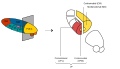
 Thalamic nuclei: MNG = Midline nuclear group AN = Anterior nuclear group MD = Medial dorsal nucleus VNG = Ventral nuclear group VA = Ventral anterior nucleus VL = Ventral lateral nucleus VPL = Ventral posterolateral nucleus VPM = Ventral posteromedial nucleus LNG = Lateral nuclear group PUL = Pulvinar MTh = Metathalamus LG = Lateral geniculate nucleus MG = Medial geniculate nucleus | |
 Thalamic nuclei | |
| Details | |
| Part of | Thalamus |
| Identifiers | |
| Latin | nuclei anteriores thalami |
| NeuroNames | 302 |
| NeuroLex ID | birnlex_1692 |
| TA98 | A14.1.08.603 |
| TA2 | 5679 |
| FMA | 62019 |
| Anatomical terms of neuroanatomy | |
The anterior nuclei of thalamus (or anterior nuclear group) are a collection of nuclei at the rostral end of the dorsal thalamus. They comprise the anteromedial, anterodorsal, and anteroventral nuclei.