The striatum or corpus striatum is a cluster of interconnected nuclei that make up the largest structure of the subcortical basal ganglia. The striatum is a critical component of the motor and reward systems; receives glutamatergic and dopaminergic inputs from different sources; and serves as the primary input to the rest of the basal ganglia.
The basal ganglia (BG) or basal nuclei are a group of subcortical nuclei found in the brains of vertebrates. In humans and other primates, differences exist, primarily in the division of the globus pallidus into external and internal regions, and in the division of the striatum. Positioned at the base of the forebrain and the top of the midbrain, they have strong connections with the cerebral cortex, thalamus, brainstem and other brain areas. The basal ganglia are associated with a variety of functions, including regulating voluntary motor movements, procedural learning, habit formation, conditional learning, eye movements, cognition, and emotion.

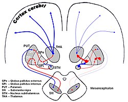
The globus pallidus (GP), also known as paleostriatum or dorsal pallidum, is a major component of the subcortical basal ganglia in the brain. It consists of two adjacent segments, one external, known in rodents simply as the globus pallidus, and one internal. It is part of the telencephalon, but retains close functional ties with the subthalamus in the diencephalon – both of which are part of the extrapyramidal motor system.

The internal capsule is a paired white matter structure, as a two-way tract, carrying ascending and descending fibers, to and from the cerebral cortex. The internal capsule is situated in the inferomedial part of each cerebral hemisphere of the brain. It carries information past the subcortical basal ganglia. As it courses it separates the caudate nucleus and the thalamus from the putamen and the globus pallidus. It also separates the caudate nucleus and the putamen in the dorsal striatum, a brain region involved in motor and reward pathways.

In the anatomy of the brain, the centromedian nucleus, also known as the centrum medianum, is a nucleus in the posterior group of the intralaminar thalamic nuclei (ITN) in the thalamus. There are two centromedian nuclei arranged bilaterally.

The thalamic reticular nucleus is part of the ventral thalamus that forms a capsule around the thalamus laterally. However, recent evidence from mice and fish question this statement and define it as a dorsal thalamic structure. It is separated from the thalamus by the external medullary lamina. Reticular nucleus cells are all GABAergic, and have discoid dendritic arbors in the plane of the nucleus.

The lentiform nucleus are the putamen (laterally) and the globus pallidus (medially), collectively. Due to their proximity, these two structures were formerly considered one, however, the two are separated by a thin layer of white matter - the external medullary lamina - and are functionally and connectionally distinct.

The subthalamus or ventral thalamus is a part of the diencephalon. Its most prominent structure is the subthalamic nucleus. The subthalamus connects to the globus pallidus, a subcortical nucleus of the basal ganglia.

The substantia innominata, also innominate substance or substantia innominata of Meynert, is a series of layers in the human brain consisting partly of gray and partly of white matter, which lies below the anterior part of the thalamus and lentiform nucleus. It is included as part of the anterior perforated substance. It is part of the basal forebrain structures and includes the nucleus basalis. A portion of the substantia innominata, below the globus pallidus is considered as part of the extended amygdala.

The basal ganglia form a major brain system in all vertebrates, but in primates there are special differentiating features. The basal ganglia include the striatum, globus pallidus, substantia nigra and subthalamic nucleus. In primates the pallidus is divided into an external and internal globus pallidus, the external globus pallidus is present in other mammals but not the internal globus pallidus. Also in primates, the dorsal striatum is divided by a large nerve tract called the internal capsule into two masses named the caudate nucleus and the putamen. These differences contribute to a complex circuitry of connections between the striatum and cortex that is specific to primates, reflecting different functions in primate cortical areas.
The mammillothalamic tract (MMT) is an efferent pathway of the mammillary bodies which project to the anterior nuclei of the thalamus. The mammillothalamic tract is part of the Papez circuit, starting and finishing in the hippocampus. The fibers of the MMT are heavily myelinated.

The ventral anterior nucleus (VA) is a nucleus in the ventral nuclear group of the thalamus. It acts with the anterior part of the ventral lateral nucleus to modify signals from the basal ganglia.
The ventral lateral nucleus (VL) is a nucleus in the ventral nuclear group of the thalamus.

The thalamic fasciculus is a component of the subthalamus (ventral thalamus). It is synonymous with field H1 of Forel. Fibers from the lenticular fasciculus (field H2 of Forel), are joined by fibers from the ansa lenticularis – different parts of the internal globus pallidus, before they enter the ventral anterior nucleus of the thalamus to form the thalamic fasciculus. The fasciculus also contains fibers from the cerebellothalamic tract, and the pallidothalamic tract.
The external globus pallidus combines with the internal globus pallidus (GPi) to form the globus pallidus, an anatomical subset of the basal ganglia. Globus pallidus means "pale globe" in Latin, indicating its appearance. The external globus pallidus is the segment of the globus pallidus that is relatively further (lateral) from the midline of the brain.
The internal globus pallidus is one of the two subcortical nuclei that provides inhibitory output in the basal ganglia, the other being the substantia nigra pars reticulata. Together with the external globus pallidus (GPe), it makes up one of the two segments of the globus pallidus, a structure that can decay with certain neurodegenerative disorders and is a target for medical and neurosurgical therapies. The GPi, along with the substantia nigra pars reticulata, comprise the primary output of the basal ganglia, with its outgoing GABAergic neurons having an inhibitory function in the thalamus, the centromedian complex and the pedunculopontine complex.
The subthalamic fasciculus is a bi-directional nerve tract that interconnects the lateral globus pallidus and subthalamic nucleus as part of the indirect pathway. The pallidal efferents are GABAergic, but also contain enkephalin. Its fibers interweave perpendicularly among the fibers of the internal capsule.
The pallidothalamic tracts are a part of the basal ganglia. They provide connectivity between the internal globus pallidus (GPi) and the thalamus, primarily the ventral anterior nucleus and the ventral lateral nucleus.

Blocq's disease was first considered by Paul Blocq (1860–1896), who described this phenomenon as the loss of memory of specialized movements causing the inability to maintain an upright posture, despite normal function of the legs in the bed. The patient is able to stand up, but as soon as the feet are on the ground, the patient cannot hold himself upright nor walk; however when lying down, the subject conserved the integrity of muscular force and the precision of movements of the lower limbs. The motivation of this study came when a fellow student Georges Marinesco (1864) and Paul published a case of parkinsonian tremor (1893) due to a tumor located in the substantia nigra.
The fields of Forel is a complex region in the posterior subthalamus, consisting of a concentrated collection of bundles of fibers. The tracts formed include the thalamic fasciculus that includes the ansa lenticularis and lenticular fasciculus, cerebellothalamic tracts, and pallidothalamic tracts. Other included fibers connect to other brain regions. These tracts are described in regions known as H fields.