| Ventral anterior nucleus | |
|---|---|
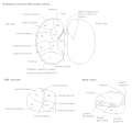
 Thalamic nuclei: MNG = Midline nuclear group AN = Anterior nuclear group MD = Medial dorsal nucleus VNG = Ventral nuclear group VA = Ventral anterior nucleus VL = Ventral lateral nucleus VPL = Ventral posterolateral nucleus VPM = Ventral posteromedial nucleus LNG = Lateral nuclear group PUL = Pulvinar MTh = Metathalamus LG = Lateral geniculate nucleus MG = Medial geniculate nucleus | |
 Thalamic nuclei | |
| Details | |
| Identifiers | |
| Latin | nucleus ventralis anterior thalami |
| NeuroNames | 334 |
| NeuroLex ID | birnlex_1232 |
| TA98 | A14.1.08.652 |
| TA2 | 5688 |
| FMA | 62184 |
| Anatomical terms of neuroanatomy | |
The ventral anterior nucleus (VA) is a nucleus in the ventral nuclear group of the thalamus. It acts with the anterior part of the ventral lateral nucleus to modify signals from the basal ganglia. [1]