Centromedian nucleus

Last updated
Centromedian nucleus
Details
Part of Intralaminar thalamic nuclei
Identifiers
Latin nucleus centromedianus thalami
Acronym(s)CM or Cm-Pf
NeuroNames 323
NeuroLex ID birnlex_805
TA98 A14.1.08.618
FMA 62165
Anatomical terms of neuroanatomy

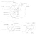
In the anatomy of the brain, the centromedian nucleus, also known as the centrum medianum, (CM or Cm-Pf) is a part of the intralaminar thalamic nuclei (ITN) in the thalamus. There are two centromedian nuclei arranged bilaterally.

Contents

In humans, it contains about 2000 neurons per cubic millimetre and has a volume of about 310 cubic millimetres with 664,000 neurons in total. [1]

Input and output

It sends nerve fibres to the subthalamic nucleus and putamen. [2] It receives nerve fibres from the cerebral cortex, vestibular nuclei, globus pallidus, superior colliculus, reticular formation, and spinothalamic tract.

Function

Its physiological role involves attention and arousal, including control of the level of cortical activity. Some frequencies of extracellular electrical stimulation of the centromedian nucleus can cause absence seizures (temporary loss of consciousness) although electrical stimulation can be of therapeutic use in intractable epilepsy and Tourette's syndrome. Specifically, centromedian nucleus has been proposed to be a target for neuromodulation-based treatment of generalized epilepsy. [3] General anaesthetics specifically suppress activity in the ILN, including the centromedian nucleus. Complete bilateral lesions of the centromedian nucleus can lead to states normally associated with brain death such as coma, death, persistent vegetative state, forms of mutism and severe delirium. Unilateral lesions can lead to unilateral thalamic neglect.

A patient with electrodes implanted into more than 50 different regions in his brain (including regions giving him orgasmic feelings) choose to self stimulate the electrode in his centromedian nucleus more than all other electrodes. The patients explanation of this: "The subject reported that he was almost able to recall a memory during this stimulation, but he could not quite grasp it. The frequent selfstimulations were an endeavor to bring this elusive memory into clear focus." [4]

Additional images

Notes and references

  1. Henderson J, Carpenter K, Cartwright H, Halliday G (2000). "Loss of thalamic intralaminar nuclei in progressive supranuclear palsy and Parkinson's disease: clinical and therapeutic implications". Brain. 123 ( Pt 7) (7): 1410–1421. doi: 10.1093/brain/123.7.1410 . PMID   10869053. Archived from the original on 2004-12-25. Retrieved 2004-09-24.
  2. Powell, T. P. S.; Cowan W. M. (1967). "The interpretation of the degenerative changes in the intralaminar nuclei of the thalamus". Journal of Neurology, Neurosurgery & Psychiatry. 30 (2): 140–153. doi:10.1136/jnnp.30.2.140. PMC   496153 . PMID   4962197.
  3. Kokkinos, Vasileios; Urban, Alexandra; Sisterson, Nathaniel D.; Li, Ningfei; Corson, Danielle; Richardson, R Mark (2020). "Responsive Neurostimulation of the Thalamus Improves Seizure Control in Idiopathic Generalized Epilepsy: A Case Report". Neurosurgery. pp. E578–E583. doi:10.1093/neuros/nyaa001. PMID   32023343.
  4. "Electrical Self-Stimulation of the Brain in 4 1". 1963. CiteSeerX   10.1.1.188.7109 .{{cite journal}}: Cite journal requires |journal= (help)

Related Research Articles

<span class="mw-page-title-main">Thalamus</span> Structure within the brain

The thalamus is a large mass of gray matter located in the dorsal part of the diencephalon. Nerve fibers project out of the thalamus to the cerebral cortex in all directions, allowing hub-like exchanges of information. It has several functions, such as the relaying of sensory signals, including motor signals to the cerebral cortex and the regulation of consciousness, sleep, and alertness.

<span class="mw-page-title-main">Trigeminal nerve</span> Cranial nerve responsible for the faces senses and motor functions

In neuroanatomy, the trigeminal nerve (lit. triplet nerve), also known as the fifth cranial nerve, cranial nerve V, or simply CN V, is a cranial nerve responsible for sensation in the face and motor functions such as biting and chewing; it is the most complex of the cranial nerves. Its name (trigeminal, from Latin tri- 'three', and -geminus 'twin') derives from each of the two nerves (one on each side of the pons) having three major branches: the ophthalmic nerve (V1), the maxillary nerve (V2), and the mandibular nerve (V3). The ophthalmic and maxillary nerves are purely sensory, whereas the mandibular nerve supplies motor as well as sensory (or "cutaneous") functions. Adding to the complexity of this nerve is that autonomic nerve fibers as well as special sensory fibers (taste) are contained within it.

<span class="mw-page-title-main">Deep brain stimulation</span> Neurosurgical treatment involving implantation of a brain pacemaker

Deep brain stimulation (DBS) is a surgical procedure that implants a neurostimulator and electrodes which sends electrical impulses to specified targets in the brain responsible for movement control. The treatment is designed for a range of movement disorders such as Parkinson's disease, essential tremor, and dystonia, as well as for certain neuropsychiatric conditions like obsessive-compulsive disorder (OCD) and epilepsy. The exact mechanisms of DBS are complex and not entirely clear, but it is known to modify brain activity in a structured way.

<span class="mw-page-title-main">Claustrum</span> Structure in the brain

The claustrum is a thin sheet of neurons and supporting glial cells, that connects to the cerebral cortex and subcortical regions including the amygdala, hippocampus and thalamus of the brain. It is located between the insular cortex laterally and the putamen medially, encased by the extreme and external capsules respectively. Blood to the claustrum is supplied by the middle cerebral artery. It is considered to be the most densely connected structure in the brain, and thus hypothesized to allow for the integration of various cortical inputs such as vision, sound and touch, into one experience. Other hypotheses suggest that the claustrum plays a role in salience processing, to direct attention towards the most behaviorally relevant stimuli amongst the background noise. The claustrum is difficult to study given the limited number of individuals with claustral lesions and the poor resolution of neuroimaging.

<span class="mw-page-title-main">Pretectal area</span> Structure in the midbrain which mediates responses to ambient light

In neuroanatomy, the pretectal area, or pretectum, is a midbrain structure composed of seven nuclei and comprises part of the subcortical visual system. Through reciprocal bilateral projections from the retina, it is involved primarily in mediating behavioral responses to acute changes in ambient light such as the pupillary light reflex, the optokinetic reflex, and temporary changes to the circadian rhythm. In addition to the pretectum's role in the visual system, the anterior pretectal nucleus has been found to mediate somatosensory and nociceptive information.

<span class="mw-page-title-main">Reticular formation</span> Spinal trigeminal nucleus

The reticular formation is a set of interconnected nuclei that are located throughout the brainstem. It is not anatomically well defined, because it includes neurons located in different parts of the brain. The neurons of the reticular formation make up a complex set of networks in the core of the brainstem that extend from the upper part of the midbrain to the lower part of the medulla oblongata. The reticular formation includes ascending pathways to the cortex in the ascending reticular activating system (ARAS) and descending pathways to the spinal cord via the reticulospinal tracts.

<span class="mw-page-title-main">Minimally conscious state</span> Disorder of Consciousness where overt signs of awareness are preserved

A minimally conscious state or MCS is a disorder of consciousness distinct from persistent vegetative state and locked-in syndrome. Unlike persistent vegetative state, patients with MCS have partial preservation of conscious awareness. MCS is a relatively new category of disorders of consciousness. The natural history and longer term outcome of MCS have not yet been thoroughly studied. The prevalence of MCS was estimated to be 9 times of PVS cases, or between 112,000 and 280,000 in the US by year 2000.

<span class="mw-page-title-main">Thalamocortical radiations</span> Neural pathways between the thalamus and cerebral cortex

In neuroanatomy, thalamocortical radiations also known as thalamocortical fibres, are the efferent fibres that project from the thalamus to distinct areas of the cerebral cortex. They form fibre bundles that emerge from the lateral surface of the thalamus.

The zona incerta (ZI) is a horizontally elongated region of gray matter in the subthalamus below the thalamus. Its connections project extensively over the brain from the cerebral cortex down into the spinal cord.

<span class="mw-page-title-main">Intralaminar thalamic nuclei</span>

The intralaminar thalamic nuclei (ITN) are collections of neurons in the internal medullary lamina of the thalamus that are generally divided in two groups as follows:

<span class="mw-page-title-main">Medullary laminae of thalamus</span>

Medullary laminae of thalamus are layers of myelinated fibres that appear on cross sections of the thalamus. They also are commonly referred to as laminae medullares thalami or medullary layers of thalamus. The specific layers are:

Recurrent thalamo-cortical resonance is an observed phenomenon of oscillatory neural activity between the thalamus and various cortical regions of the brain. It is proposed by Rodolfo Llinas and others as a theory for the integration of sensory information into the whole of perception in the brain. Thalamocortical oscillation is proposed to be a mechanism of synchronization between different cortical regions of the brain, a process known as temporal binding. This is possible through the existence of thalamocortical networks, groupings of thalamic and cortical cells that exhibit oscillatory properties.

<span class="mw-page-title-main">Internal globus pallidus</span>

The internal globus pallidus and the external globus pallidus (GPe) make up the globus pallidus. The GPi is one of the output nuclei of the basal ganglia. The GABAergic neurons of the GPi send their axons to the ventral anterior nucleus (VA) and the ventral lateral nucleus (VL) in the dorsal thalamus, to the centromedian complex, and to the pedunculopontine complex.

Ablative brain surgery is the surgical ablation by various methods of brain tissue to treat neurological or psychological disorders. The word "Ablation" stems from the Latin word Ablatus meaning "carried away". In most cases, however, ablative brain surgery does not involve removing brain tissue, but rather destroying tissue and leaving it in place. The lesions it causes are irreversible. There are some target nuclei for ablative surgery and deep brain stimulation. Those nuclei are the motor thalamus, the globus pallidus, and the subthalamic nucleus.

The cerebellothalamic tract or the tractus cerebellothalamicus, is part of the superior cerebellar peduncle. It originates in the cerebellar nuclei, crosses completely in the decussation of the superior cerebellar peduncle, bypasses the red nucleus, and terminates in posterior division of ventral lateral nucleus of thalamus. The ventrolateral nucleus has different divisions and distinct connections, mostly with frontal and parietal lobes. The primary motor cortex and premotor cortex get information from the ventrolateral nucleus projections originating in the interposed nucleus and dentate nuclei. Other dentate nucleus projections via thalamic pathway transmit information to prefrontal cortex and posterior parietal cortex. The cerebellum sends thalamocortical projections and in addition may also send connections from the thalamus to association areas serving cognitive and affective functions.

<span class="mw-page-title-main">Basal ganglia disease</span> Group of physical problems resulting from basal ganglia dysfunction

Basal ganglia disease is a group of physical problems that occur when the group of nuclei in the brain known as the basal ganglia fail to properly suppress unwanted movements or to properly prime upper motor neuron circuits to initiate motor function. Research indicates that increased output of the basal ganglia inhibits thalamocortical projection neurons. Proper activation or deactivation of these neurons is an integral component for proper movement. If something causes too much basal ganglia output, then the ventral anterior (VA) and ventral lateral (VL) thalamocortical projection neurons become too inhibited, and one cannot initiate voluntary movement. These disorders are known as hypokinetic disorders. However, a disorder leading to abnormally low output of the basal ganglia leads to reduced inhibition, and thus excitation, of the thalamocortical projection neurons which synapse onto the cortex. This situation leads to an inability to suppress unwanted movements. These disorders are known as hyperkinetic disorders.

The parabrachial nuclei, also known as the parabrachial complex, are a group of nuclei in the dorsolateral pons that surrounds the superior cerebellar peduncle as it enters the brainstem from the cerebellum. They are named from the Latin term for the superior cerebellar peduncle, the brachium conjunctivum. In the human brain, the expansion of the superior cerebellar peduncle expands the parabrachial nuclei, which form a thin strip of grey matter over most of the peduncle. The parabrachial nuclei are typically divided along the lines suggested by Baxter and Olszewski in humans, into a medial parabrachial nucleus and lateral parabrachial nucleus. These have in turn been subdivided into a dozen subnuclei: the superior, dorsal, ventral, internal, external and extreme lateral subnuclei; the lateral crescent and subparabrachial nucleus along the ventrolateral margin of the lateral parabrachial complex; and the medial and external medial subnuclei

Neuromodulation is "the alteration of nerve activity through targeted delivery of a stimulus, such as electrical stimulation or chemical agents, to specific neurological sites in the body". It is carried out to normalize – or modulate – nervous tissue function. Neuromodulation is an evolving therapy that can involve a range of electromagnetic stimuli such as a magnetic field (rTMS), an electric current, or a drug instilled directly in the subdural space. Emerging applications involve targeted introduction of genes or gene regulators and light (optogenetics), and by 2014, these had been at minimum demonstrated in mammalian models, or first-in-human data had been acquired. The most clinical experience has been with electrical stimulation.

In the human brain, the central lateral nucleus is a part of the anterior intralaminar nucleus in the thalamus. The intralaminar nuclei project to many different regions of the brain, The thalamus acts generally as a relay point for the brain for other areas of the brain to link to. The central lateral nucleus acts as a vital role in consciousness. This area of the brain also affects conditioned emotional responses, such as fear conditioning.