Grey matter

Last updated

Grey matter
Spinal nerve.svg
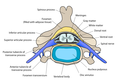
The formation of the spinal nerve from the dorsal and ventral roots (with grey matter labelled at centre right).
Grey matter and white matter - very high mag.jpg
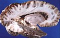
Micrograph showing grey matter, with the characteristic neuronal cell bodies (dark shade of pink), and white matter with its characteristic fine meshwork-like appearance (left of image; lighter shade of pink). HPS stain.
Details
Identifiers
Latin substantia grisea
MeSH D066128
TA98 A14.1.00.002
A14.1.02.020
A14.1.04.201
A14.1.05.201
A14.1.05.401
A14.1.06.301
TA2 5365
FMA 67242
Anatomical terminology

Grey matter, or brain matter in American English, is a major component of the central nervous system, consisting of neuronal cell bodies, neuropil (dendrites and unmyelinated axons), glial cells (astrocytes and oligodendrocytes), synapses, and capillaries. Grey matter is distinguished from white matter in that it contains numerous cell bodies and relatively few myelinated axons, while white matter contains relatively few cell bodies and is composed chiefly of long-range myelinated axons. [1] The colour difference arises mainly from the whiteness of myelin. In living tissue, grey matter actually has a very light grey colour with yellowish or pinkish hues, which come from capillary blood vessels and neuronal cell bodies. [2]

Contents

Structure

Grey matter refers to unmyelinated neurons and other cells of the central nervous system. It is present in the brain, brainstem and cerebellum, and present throughout the spinal cord.

Grey matter is distributed at the surface of the cerebral hemispheres (cerebral cortex) and of the cerebellum (cerebellar cortex), as well as in the depths of the cerebrum (the thalamus; hypothalamus; subthalamus, basal gangliaputamen, globus pallidus and nucleus accumbens; as well as the septal nuclei), cerebellum (deep cerebellar nuclei – the dentate nuclei, globose nucleus, emboliform nucleus, and fastigial nucleus), and brainstem (the substantia nigra, red nucleus, olivary nuclei, and cranial nerve nuclei).

Grey matter in the spinal cord is known as the grey column which travels down the spinal cord distributed in three grey columns that are presented in an "H" shape. The forward-facing column is the anterior grey column, the rear-facing one is the posterior grey column and the interlinking one is the lateral grey column. The grey matter on the left and right side is connected by the grey commissure. The grey matter in the spinal cord consists of interneurons, as well as the cell bodies of projection neurons.

Grey matter undergoes development and growth throughout childhood and adolescence. [3] Recent studies using cross-sectional neuroimaging have shown that by around the age of 8 the volume of grey matter begins to decrease. [4] However, the density of grey matter appears to increase as a child develops into early adulthood. [4] Males tend to exhibit grey matter of increased volume but lower density than that of females. [5]

Function

Grey matter contains most of the brain's neuronal cell bodies. [6] The grey matter includes regions of the brain involved in muscle control, and sensory perception such as seeing and hearing, memory, emotions, speech, decision-making, and self-control.

The grey matter in the spinal cord is split into three grey columns:

The grey matter of the spinal cord can be divided into different layers, called Rexed laminae. These describe, in general, the purpose of the cells within the grey matter of the spinal cord at a particular location.

Clinical significance

High alcohol consumption has been correlated with significant reductions in grey matter volume. [7] [8] Short-term cannabis use (30 days) is not correlated with changes in white or grey matter. [9] However, several cross-sectional studies have shown that repeated long-term cannabis use is associated with smaller grey matter volumes in the hippocampus, amygdala, medial temporal cortex, and prefrontal cortex, with increased grey matter volume in the cerebellum. [10] [11] [12] Long-term cannabis use is also associated with alterations in white matter integrity in an age-dependent manner, [13] with heavy cannabis use during adolescence and early adulthood associated with the greatest amount of change. [14]

Meditation has been shown to change grey matter structure. [15] [16] [17] [18] [19]

Habitual playing of action video games has been reported to promote a reduction of grey matter in the hippocampus while 3D platformer games have been reported to increase grey matter in the hippocampus. [20] [21] [22]

Women and men with equivalent IQ scores have differing proportions of grey to white matter in cortical brain regions associated with intelligence. [23]

Pregnancy renders substantial changes in brain structure, primarily reductions in grey matter volume in regions subserving social cognition. The grey matter reductions endured for at least 2 years post-pregnancy. [24] The profile of brain changes is comparable to that taking place during adolescence, a hormonally similar transitional period of life. [25]

History

Etymology

In the current edition [26] of the official Latin nomenclature, Terminologia Anatomica , substantia grisea is used for English grey matter. The adjective grisea for grey is however not attested in classical Latin. [27] The adjective grisea is derived from the French word for grey, gris. [27] Alternative designations like substantia cana [28] and substantia cinerea [29] are being used alternatively. The adjective cana, attested in classical Latin, [30] can mean grey, [27] or greyish white. [31] The classical Latin cinerea means ash-coloured. [30]

Additional images

See also

Related Research Articles

<span class="mw-page-title-main">Central nervous system</span> Brain and spinal cord

The central nervous system (CNS) is the part of the nervous system consisting of the brain and spinal cord, the retina and optic nerve, and the olfactory nerve and epithelia. The CNS is so named because the brain integrates the received information and coordinates and influences the activity of all parts of the bodies of bilaterally symmetric and triploblastic animals—that is, all multicellular animals except sponges and diploblasts. It is a structure composed of nervous tissue positioned along the rostral to caudal axis of the body and may have an enlarged section at the rostral end which is a brain. Only arthropods, cephalopods and vertebrates have a true brain, though precursor structures exist in onychophorans, gastropods and lancelets.

In physiology, nociception, also nocioception; from Latin nocere 'to harm/hurt') is the sensory nervous system's process of encoding noxious stimuli. It deals with a series of events and processes required for an organism to receive a painful stimulus, convert it to a molecular signal, and recognize and characterize the signal to trigger an appropriate defensive response.

<span class="mw-page-title-main">White matter</span> Areas of myelinated axons in the brain

White matter refers to areas of the central nervous system (CNS) that are mainly made up of myelinated axons, also called tracts. Long thought to be passive tissue, white matter affects learning and brain functions, modulating the distribution of action potentials, acting as a relay and coordinating communication between different brain regions.

<span class="mw-page-title-main">Cerebellum</span> Structure at the rear of the vertebrate brain, beneath the cerebrum

The cerebellum is a major feature of the hindbrain of all vertebrates. Although usually smaller than the cerebrum, in some animals such as the mormyrid fishes it may be as large as it or even larger. In humans, the cerebellum plays an important role in motor control. It may also be involved in some cognitive functions such as attention and language as well as emotional control such as regulating fear and pleasure responses, but its movement-related functions are the most solidly established. The human cerebellum does not initiate movement, but contributes to coordination, precision, and accurate timing: it receives input from sensory systems of the spinal cord and from other parts of the brain, and integrates these inputs to fine-tune motor activity. Cerebellar damage produces disorders in fine movement, equilibrium, posture, and motor learning in humans.

<span class="mw-page-title-main">Brainstem</span> Posterior part of the brain, adjoining and structurally continuous

The brainstem is the stalk-like part of the brain that interconnects the cerebrum and diencephalon with the spinal cord. In the human brain, the brainstem is composed of the midbrain, the pons, and the medulla oblongata. The midbrain is continuous with the thalamus of the diencephalon through the tentorial notch.

In neuroanatomy, a nucleus is a cluster of neurons in the central nervous system, located deep within the cerebral hemispheres and brainstem. The neurons in one nucleus usually have roughly similar connections and functions. Nuclei are connected to other nuclei by tracts, the bundles (fascicles) of axons extending from the cell bodies. A nucleus is one of the two most common forms of nerve cell organization, the other being layered structures such as the cerebral cortex or cerebellar cortex. In anatomical sections, a nucleus shows up as a region of gray matter, often bordered by white matter. The vertebrate brain contains hundreds of distinguishable nuclei, varying widely in shape and size. A nucleus may itself have a complex internal structure, with multiple types of neurons arranged in clumps (subnuclei) or layers.

<span class="mw-page-title-main">Neural pathway</span> Connection formed between neurons that allows neurotransmission

In neuroanatomy, a neural pathway is the connection formed by axons that project from neurons to make synapses onto neurons in another location, to enable neurotransmission. Neurons are connected by a single axon, or by a bundle of axons known as a nerve tract, or fasciculus. Shorter neural pathways are found within grey matter in the brain, whereas longer projections, made up of myelinated axons, constitute white matter.

<span class="mw-page-title-main">Grey column</span>

The grey column refers to a somewhat ridge-shaped mass of grey matter in the spinal cord. This presents as three columns: the anterior grey column, the posterior grey column, and the lateral grey column, all of which are visible in cross-section of the spinal cord.

<span class="mw-page-title-main">Pyramidal tracts</span> The corticobulbar tract and the corticospinal tract

The pyramidal tracts include both the corticobulbar tract and the corticospinal tract. These are aggregations of efferent nerve fibers from the upper motor neurons that travel from the cerebral cortex and terminate either in the brainstem (corticobulbar) or spinal cord (corticospinal) and are involved in the control of motor functions of the body.

<span class="mw-page-title-main">Spinothalamic tract</span> Sensory pathway from the skin to the thalamus

The spinothalamic tract is a part of the anterolateral system or the ventrolateral system, a sensory pathway to the thalamus. From the ventral posterolateral nucleus in the thalamus, sensory information is relayed upward to the somatosensory cortex of the postcentral gyrus.

<span class="mw-page-title-main">Dorsal column–medial lemniscus pathway</span> Sensory spinal pathway

The dorsal column–medial lemniscus pathway (DCML) is a sensory pathway of the central nervous system that conveys sensations of fine touch, vibration, two-point discrimination, and proprioception from the skin and joints. It transmits information from the body to the primary somatosensory cortex in the postcentral gyrus of the parietal lobe of the brain. The pathway receives information from sensory receptors throughout the body, and carries this in nerve tracts in the white matter of the dorsal column of the spinal cord to the medulla, where it is continued in the medial lemniscus, on to the thalamus and relayed from there through the internal capsule and transmitted to the somatosensory cortex. The name dorsal-column medial lemniscus comes from the two structures that carry the sensory information: the dorsal columns of the spinal cord, and the medial lemniscus in the brainstem.

<span class="mw-page-title-main">Locus coeruleus</span> Stress and panic response centre

The locus coeruleus (LC), also spelled locus caeruleus or locus ceruleus, is a nucleus in the pons of the brainstem involved with physiological responses to stress and panic. It is a part of the reticular activating system.

<span class="mw-page-title-main">Reticular formation</span> Spinal trigeminal nucleus

The reticular formation is a set of interconnected nuclei that are located in the brainstem, hypothalamus, and other regions. It is not anatomically well defined, because it includes neurons located in different parts of the brain. The neurons of the reticular formation make up a complex set of networks in the core of the brainstem that extend from the upper part of the midbrain to the lower part of the medulla oblongata. The reticular formation includes ascending pathways to the cortex in the ascending reticular activating system (ARAS) and descending pathways to the spinal cord via the reticulospinal tracts.

<span class="mw-page-title-main">Spinocerebellar tract</span> Nerve tract in humans

The spinocerebellar tract is a nerve tract originating in the spinal cord and terminating in the same side (ipsilateral) of the cerebellum.

The zona incerta (ZI) is a horizontally elongated region of gray matter in the subthalamus below the thalamus. Its connections project extensively over the brain from the cerebral cortex down into the spinal cord.

<span class="mw-page-title-main">Nucleus raphe magnus</span>

The nucleus raphe magnus is one of the seven raphe nuclei. It is situated in the pons in the brainstem, just rostral to the nucleus raphe obscurus.

<span class="mw-page-title-main">Posterolateral tract</span>

The posterolateral tract is a small strand situated in relation to the tip of the posterior column close to the entrance of the posterior nerve roots. It is present throughout the spinal cord, and is most developed in the upper cervical regions.

<span class="mw-page-title-main">Spinal cord</span> Long, tubular central nervous system structure in the vertebral column

The spinal cord is a long, thin, tubular structure made up of nervous tissue that extends from the medulla oblongata in the brainstem to the lumbar region of the vertebral column (backbone) of vertebrate animals. The center of the spinal cord is hollow and contains a structure called the central canal, which contains cerebrospinal fluid. The spinal cord is also covered by meninges and enclosed by the neural arches. Together, the brain and spinal cord make up the central nervous system.

<span class="mw-page-title-main">Anatomy of the cerebellum</span> Structures in the cerebellum, a part of the brain

The anatomy of the cerebellum can be viewed at three levels. At the level of gross anatomy, the cerebellum consists of a tightly folded and crumpled layer of cortex, with white matter underneath, several deep nuclei embedded in the white matter, and a fluid-filled ventricle in the middle. At the intermediate level, the cerebellum and its auxiliary structures can be broken down into several hundred or thousand independently functioning modules or compartments known as microzones. At the microscopic level, each module consists of the same small set of neuronal elements, laid out with a highly stereotyped geometry.

The parabrachial nuclei, also known as the parabrachial complex, are a group of nuclei in the dorsolateral pons that surrounds the superior cerebellar peduncle as it enters the brainstem from the cerebellum. They are named from the Latin term for the superior cerebellar peduncle, the brachium conjunctivum. In the human brain, the expansion of the superior cerebellar peduncle expands the parabrachial nuclei, which form a thin strip of grey matter over most of the peduncle. The parabrachial nuclei are typically divided along the lines suggested by Baxter and Olszewski in humans, into a medial parabrachial nucleus and lateral parabrachial nucleus. These have in turn been subdivided into a dozen subnuclei: the superior, dorsal, ventral, internal, external and extreme lateral subnuclei; the lateral crescent and subparabrachial nucleus along the ventrolateral margin of the lateral parabrachial complex; and the medial and external medial subnuclei

References

  1. Purves D, Augustine GJ, Fitzpatrick D, Hall WC, LaMantia AS, McNamara JO, White LE (2008). Neuroscience (4th ed.). Sinauer Associates. pp. 15–16. ISBN   978-0-87893-697-7.
  2. Kolb B, Whishaw IQ (2003). Fundamentals of human neuropsychology (5th ed.). New York: Worth Publishing. p. 49. ISBN   978-0-7167-5300-1.
  3. Sowell ER, Thompson PM, Tessner KD, Toga AW (November 2001). "Mapping continued brain growth and gray matter density reduction in dorsal frontal cortex: Inverse relationships during postadolescent brain maturation". The Journal of Neuroscience. 21 (22): 8819–29. doi:10.1523/JNEUROSCI.21-22-08819.2001. PMC   6762261 . PMID   11698594.
  4. 1 2 Gennatas ED, Avants BB, Wolf DH, Satterthwaite TD, Ruparel K, Ciric R, Hakonarson H, Gur RE, Gur RC (May 2017). "Age-Related Effects and Sex Differences in Gray Matter Density, Volume, Mass, and Cortical Thickness from Childhood to Young Adulthood". The Journal of Neuroscience. 37 (20): 5065–5073. doi:10.1523/JNEUROSCI.3550-16.2017. PMC   5444192 . PMID   28432144.
  5. Luders, Eileen; Gaser, Christian; Narr, Katherine L.; Toga, Arthur W. (11 November 2009). "Why Sex Matters: Brain Size Independent Differences in Gray Matter Distributions between Men and Women". The Journal of Neuroscience. 29 (45): 14265–14270. doi:10.1523/JNEUROSCI.2261-09.2009. PMC   3110817 . PMID   19906974.
  6. Miller AK, Alston RL, Corsellis JA (1980). "Variation with age in the volumes of grey and white matter in the cerebral hemispheres of man: measurements with an image analyser". Neuropathology and Applied Neurobiology. 6 (2): 119–32. doi:10.1111/j.1365-2990.1980.tb00283.x. PMID   7374914. S2CID   23201991.
  7. Yang X, Tian F, Zhang H, Zeng J, Chen T, Wang S, Jia Z, Gong Q (July 2016). "Cortical and subcortical gray matter shrinkage in alcohol-use disorders: a voxel-based meta-analysis". Neuroscience and Biobehavioral Reviews. 66: 92–103. doi:10.1016/j.neubiorev.2016.03.034. PMID   27108216. S2CID   19928689.
  8. Xiao P, Dai Z, Zhong J, Zhu Y, Shi H, Pan P (August 2015). "Regional gray matter deficits in alcohol dependence: A meta-analysis of voxel-based morphometry studies". Drug and Alcohol Dependence. 153: 22–8. doi:10.1016/j.drugalcdep.2015.05.030. PMID   26072220.
  9. Thayer RE, YorkWilliams S, Karoly HC, Sabbineni A, Ewing SF, Bryan AD, Hutchison KE (December 2017). "Structural neuroimaging correlates of alcohol and cannabis use in adolescents and adults". Addiction. 112 (12): 2144–2154. doi:10.1111/add.13923. PMC   5673530 . PMID   28646566.
  10. Lorenzetti V, Lubman DI, Whittle S, Solowij N, Yücel M (September 2010). "Structural MRI findings in long-term cannabis users: what do we know?". Substance Use & Misuse. 45 (11): 1787–808. doi:10.3109/10826084.2010.482443. PMID   20590400. S2CID   22127231.
  11. Matochik JA, Eldreth DA, Cadet JL, Bolla KI (January 2005). "Altered brain tissue composition in heavy marijuana users". Drug and Alcohol Dependence. 77 (1): 23–30. doi:10.1016/j.drugalcdep.2004.06.011. PMID   15607838.
  12. Yücel M, Solowij N, Respondek C, Whittle S, Fornito A, Pantelis C, Lubman DI (June 2008). "Regional brain abnormalities associated with long-term heavy cannabis use". Archives of General Psychiatry. 65 (6): 694–701. doi: 10.1001/archpsyc.65.6.694 . PMID   18519827.
  13. Jakabek D, Yücel M, Lorenzetti V, Solowij N (October 2016). "An MRI study of white matter tract integrity in regular cannabis users: effects of cannabis use and age". Psychopharmacology. 233 (19–20): 3627–37. doi:10.1007/s00213-016-4398-3. PMID   27503373. S2CID   5968884.
  14. Becker MP, Collins PF, Lim KO, Muetzel RL, Luciana M (December 2015). "Longitudinal changes in white matter microstructure after heavy cannabis use". Developmental Cognitive Neuroscience. 16: 23–35. doi:10.1016/j.dcn.2015.10.004. PMC   4691379 . PMID   26602958.
  15. Kurth F, Luders E, Wu B, Black DS (2014). "Brain Gray Matter Changes Associated with Mindfulness Meditation in Older Adults: An Exploratory Pilot Study using Voxel-based Morphometry". Neuro. 1 (1): 23–26. doi:10.17140/NOJ-1-106. PMC   4306280 . PMID   25632405.
  16. Hölzel BK, Carmody J, Vangel M, Congleton C, Yerramsetti SM, Gard T, Lazar SW (January 2011). "Mindfulness practice leads to increases in regional brain gray matter density". Psychiatry Research. 191 (1): 36–43. doi:10.1016/j.pscychresns.2010.08.006. PMC   3004979 . PMID   21071182.
  17. Kurth F, MacKenzie-Graham A, Toga AW, Luders E (January 2015). "Shifting brain asymmetry: the link between meditation and structural lateralization". Social Cognitive and Affective Neuroscience. 10 (1): 55–61. doi:10.1093/scan/nsu029. PMC   4994843 . PMID   24643652.
  18. Fox KC, Nijeboer S, Dixon ML, Floman JL, Ellamil M, Rumak SP, Sedlmeier P, Christoff K (June 2014). "Is meditation associated with altered brain structure? A systematic review and meta-analysis of morphometric neuroimaging in meditation practitioners". Neuroscience and Biobehavioral Reviews. 43: 48–73. doi:10.1016/j.neubiorev.2014.03.016. PMID   24705269. S2CID   207090878.
  19. Hölzel BK, Carmody J, Evans KC, Hoge EA, Dusek JA, Morgan L, Pitman RK, Lazar SW (March 2010). "Stress reduction correlates with structural changes in the amygdala". Social Cognitive and Affective Neuroscience. 5 (1): 11–7. doi:10.1093/scan/nsp034. PMC   2840837 . PMID   19776221.
  20. West, Greg L.; Drisdelle, Brandi Lee; Konishi, Kyoko; Jackson, Jonathan; Jolicoeur, Pierre; Bohbot, Veronique D. (7 June 2015). "Habitual action video game playing is associated with caudate nucleus-dependent navigational strategies". Proceedings of the Royal Society B: Biological Sciences. 282 (1808): 20142952. doi:10.1098/rspb.2014.2952. PMC   4455792 . PMID   25994669.
  21. Collins K (10 August 2017). "Video games can either grow or shrink part of your brain, depending on how you play". qz.com. Archived from the original on 14 April 2018. Retrieved 5 May 2018.
  22. West GL, Zendel BR, Konishi K, Benady-Chorney J, Bohbot VD, Peretz I, Belleville S (5 May 2018). "Playing Super Mario 64 increases hippocampal grey matter in older adults". PLOS ONE. 12 (12): e0187779. doi: 10.1371/journal.pone.0187779 . PMC   5718432 . PMID   29211727.
  23. Haier RJ, Jung RE, Yeo RA, Head K, Alkire MT (March 2005). "The neuroanatomy of general intelligence: sex matters". NeuroImage. 25 (1): 320–7. doi:10.1016/j.neuroimage.2004.11.019. PMID   15734366. S2CID   4127512.
  24. Hoekzema E, Barba-Müller E, Pozzobon C, Picado M, Lucco F, García-García D, Soliva JC, Tobeña A, Desco M, Crone EA, Ballesteros A, Carmona S, Vilarroya O (February 2017). "Pregnancy leads to long-lasting changes in human brain structure". Nature Neuroscience. 20 (2): 287–296. doi:10.1038/nn.4458. hdl: 1887/57549 . PMID   27991897. S2CID   4113669.
  25. Carmona S, Martínez-García M, Paternina-Die M, Barba-Müller E, Wierenga LM, Alemán-Gómez Y, Cortizo R, Pozzobon C, Picado M, Lucco F, García-García D, Soliva JC, Tobeña A, Peper JS, Crone EA, Ballesteros A, Vilarroya O, Desco M, Hoekzema E (January 2019). "Pregnancy and adolescence entail similar neuroanatomical adaptations: A comparative analysis of cerebralmorphometric changes". Hum Brain Mapp. 40 (7): 2143–2152. doi: 10.1002/hbm.24513 . PMC   6865685 . PMID   30663172.
  26. Federative Committee on Anatomical Terminology (FCAT) (1998). Terminologia Anatomica. Stuttgart: Thieme[ page needed ]
  27. 1 2 3 Triepel H (1910). Die anatomischen Namen. Ihre Ableitung und Aussprache. Mit einem Anhang: Biographische Notizen (3rd ed.). Wiesbaden: Verlag J.F. Bergmann.[ page needed ]
  28. Triepel H (1910). Nomina Anatomica. Mit Unterstützung von Fachphilologen. Wiesbaden: Verlag J.F. Bergmann.[ page needed ]
  29. Schreger CH (1805). "Synonymia anatomica. Synonymik der anatomischen Nomenclatur". In Fürth (ed.). Bureau für Literatur.[ page needed ]
  30. 1 2 Lewis CT, Short C (1879). A Latin dictionary founded on Andrews' edition of Freund's Latin dictionary. Oxford: Clarendon Press.[ page needed ]
  31. Stearn WT (1983). Charles D (ed.). Botanical Latin. History, grammar, syntax, terminology and vocabulary (3rd ed.). London: Newton Abbot.[ page needed ]