Parietal lobe

Last updated
Parietal lobe
Principal fissures and lobes of the cerebrum viewed laterally. (Parietal lobe is shown in yellow)
Gray726 parietal lobe.png
Lateral surface of left cerebral hemisphere, viewed from the side. (Parietal lobe is shown in orange.)
Details
Pronunciation /pəˈr.ə.tl/ ) [1]
Part of Cerebrum
Artery Anterior cerebral
Middle cerebral
Vein Superior sagittal sinus
Identifiers
Latin lobus parietalis
MeSH D010296
NeuroNames 95
NeuroLex ID birnlex_1148
TA98 A14.1.09.123
TA2 5467
FMA 61826
Anatomical terms of neuroanatomy

The parietal lobe is one of the four major lobes of the cerebral cortex in the brain of mammals. The parietal lobe is positioned above the temporal lobe and behind the frontal lobe and central sulcus.

Contents

The parietal lobe integrates sensory information among various modalities, including spatial sense and navigation (proprioception), the main sensory receptive area for the sense of touch in the somatosensory cortex which is just posterior to the central sulcus in the postcentral gyrus, [2] and the dorsal stream of the visual system. The major sensory inputs from the skin (touch, temperature, and pain receptors), relay through the thalamus to the parietal lobe.

Several areas of the parietal lobe are important in language processing. The somatosensory cortex can be illustrated as a distorted figure – the cortical homunculus [3] (Latin: "little man") in which the body parts are rendered according to how much of the somatosensory cortex is devoted to them. [4] The superior parietal lobule and inferior parietal lobule are the primary areas of body or spatial awareness. A lesion commonly in the right superior or inferior parietal lobule leads to hemineglect.

The name comes from the parietal bone, which is named from the Latin paries-, meaning "wall".
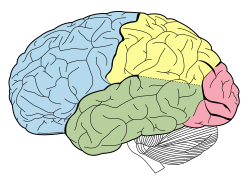
Structure

Animation. Parietal lobe (red) of left cerebral hemisphere. Parietal lobe animation small.gif
Animation. Parietal lobe (red) of left cerebral hemisphere.

The parietal lobe is defined by three anatomical boundaries: The central sulcus separates the parietal lobe from the frontal lobe; the parieto-occipital sulcus separates the parietal and occipital lobes; the lateral sulcus (sylvian fissure) is the most lateral boundary, separating it from the temporal lobe; and the longitudinal fissure divides the two hemispheres. Within each hemisphere, the somatosensory cortex represents the skin area on the contralateral surface of the body. [4]

Immediately posterior to the central sulcus, and the most anterior part of the parietal lobe, is the postcentral gyrus (Brodmann area 3), the primary somatosensory cortical area. Separating this from the posterior parietal cortex is the postcentral sulcus.

The posterior parietal cortex can be subdivided into the superior parietal lobule (Brodmann areas 5 + 7) and the inferior parietal lobule (39 + 40), separated by the intraparietal sulcus (IPS). The intraparietal sulcus and adjacent gyri are essential in guidance of limb and eye movement, and—based on cytoarchitectural and functional differences—is further divided into medial (MIP), lateral (LIP), ventral (VIP), and anterior (AIP) areas.

Function

Functions of the parietal lobe include:

The parietal lobe plays important roles in integrating sensory information from various parts of the body, knowledge of numbers and their relations, [5] and in the manipulation of objects. Its function also includes processing information relating to the sense of touch. [6] Portions of the parietal lobe are involved with visuospatial processing. [7] Although multisensory in nature, the posterior parietal cortex is often referred to by vision scientists as the dorsal stream of vision (as opposed to the ventral stream in the temporal lobe). This dorsal stream has been called both the "where" stream (as in spatial vision) [8] and the "how" stream (as in vision for action). [9] The posterior parietal cortex (PPC) receives somatosensory and visual input, which then, through motor signals, controls movement of the arm, hand, and eyes. [10]

Various studies in the 1990s found that different regions of the posterior parietal cortex in macaques represent different parts of space.

More recent fMRI studies have shown that humans have similar functional regions in and around the intraparietal sulcus and parietal-occipital junction. [18] The human "parietal eye fields" and "parietal reach region", equivalent to LIP and MIP in the monkey, also appear to be organized in gaze-centered coordinates so that their goal-related activity is "remapped" when the eyes move. [19]

Emerging evidence has linked processing in the inferior parietal lobe to declarative memory. Bilateral damage to this brain region does not cause amnesia however the strength of memory is diminished, details of complex events become harder to retrieve, and subjective confidence in memory is very low. [20] [21] [22] This has been interpreted as reflecting either deficits in internal attention, [23] deficits in subjective memory states, [22] or problems with the computation that allows evidence to accumulate, thus allowing decisions to be made about internal representations. [20]

Clinical significance

Features of parietal lobe lesions are as follows:

Damage to this lobe in the right hemisphere results in the loss of imagery, visualization of spatial relationships and neglect of left-side space and left side of the body. Even drawings may be neglected on the left side. Damage to this lobe in the left hemisphere will result in problems in mathematics, long reading, writing, and understanding symbols. The parietal association cortex enables individuals to read, write, and solve mathematical problems. The sensory inputs from the right side of the body go to the left side of the brain and vice versa.

The syndrome of hemispatial neglect is usually associated with large deficits of attention of the non-dominant hemisphere. Optic ataxia is associated with difficulties reaching toward objects in the visual field opposite to the side of the parietal damage. Some aspects of optic ataxia have been explained in terms of the functional organization described above.

Apraxia is a disorder of motor control which can be referred neither to "elemental" motor deficits nor to general cognitive impairment. The concept of apraxia was shaped by Hugo Liepmann about a hundred years ago.[ clarification needed ] [24] [25] Apraxia is predominantly a symptom of left brain damage, but some symptoms of apraxia can also occur after right brain damage. [26]

Amorphosynthesis is a loss of perception on one side of the body caused by a lesion in the parietal lobe. Usually, left-sided lesions cause agnosia, a full-body loss of perception, while right-sided lesions cause lack of recognition of the person's left side and extrapersonal space. The term amorphosynthesis was coined by D. Denny-Brown to describe patients he studied in the 1950s. [27]

Can also result in sensory impairment where one of the affected person's senses (sight, hearing, smell, touch, taste and spatial awareness) is no longer normal.[ clarification needed ] [28] [29]

See also

Related Research Articles

<span class="mw-page-title-main">Cerebral cortex</span> Outer layer of the cerebrum of the mammalian brain

The cerebral cortex, also known as the cerebral mantle, is the outer layer of neural tissue of the cerebrum of the brain in humans and other mammals. The cerebral cortex mostly consists of the six-layered neocortex, with just 10% consisting of the allocortex. It is separated into two cortices, by the longitudinal fissure that divides the cerebrum into the left and right cerebral hemispheres. The two hemispheres are joined beneath the cortex by the corpus callosum. The cerebral cortex is the largest site of neural integration in the central nervous system. It plays a key role in attention, perception, awareness, thought, memory, language, and consciousness. The cerebral cortex is part of the brain responsible for cognition.

<span class="mw-page-title-main">Cingulate cortex</span> Part of the brain within the cerebral cortex

The cingulate cortex is a part of the brain situated in the medial aspect of the cerebral cortex. The cingulate cortex includes the entire cingulate gyrus, which lies immediately above the corpus callosum, and the continuation of this in the cingulate sulcus. The cingulate cortex is usually considered part of the limbic lobe.

<span class="mw-page-title-main">Precuneus</span> Region of the parietal lobe of the brain

In neuroanatomy, the precuneus is the portion of the superior parietal lobule on the medial surface of each brain hemisphere. It is located in front of the cuneus. The precuneus is bounded in front by the marginal branch of the cingulate sulcus, at the rear by the parieto-occipital sulcus, and underneath by the subparietal sulcus. It is involved with episodic memory, visuospatial processing, reflections upon self, and aspects of consciousness.

<span class="mw-page-title-main">Dorsal column–medial lemniscus pathway</span> Sensory spinal pathway

The dorsal column–medial lemniscus pathway (DCML) is a sensory pathway of the central nervous system that conveys sensations of fine touch, vibration, two-point discrimination, and proprioception from the skin and joints. It transmits information from the body to the primary somatosensory cortex in the postcentral gyrus of the parietal lobe of the brain. The pathway receives information from sensory receptors throughout the body, and carries this in nerve tracts in the white matter of the dorsal column of the spinal cord to the medulla, where it is continued in the medial lemniscus, on to the thalamus and relayed from there through the internal capsule and transmitted to the somatosensory cortex. The name dorsal-column medial lemniscus comes from the two structures that carry the sensory information: the dorsal columns of the spinal cord, and the medial lemniscus in the brainstem.

Multisensory integration, also known as multimodal integration, is the study of how information from the different sensory modalities may be integrated by the nervous system. A coherent representation of objects combining modalities enables animals to have meaningful perceptual experiences. Indeed, multisensory integration is central to adaptive behavior because it allows animals to perceive a world of coherent perceptual entities. Multisensory integration also deals with how different sensory modalities interact with one another and alter each other's processing.

<span class="mw-page-title-main">Supramarginal gyrus</span> Gyrus of the parietal lobe of the brain

The supramarginal gyrus is a portion of the parietal lobe. This area of the brain is also known as Brodmann area 40 based on the brain map created by Korbinian Brodmann to define the structures in the cerebral cortex. It is probably involved with language perception and processing, and lesions in it may cause receptive aphasia.

<span class="mw-page-title-main">Angular gyrus</span> Gyrus of the parietal lobe of the brain

The angular gyrus is a region of the brain lying mainly in the posteroinferior region of the parietal lobe, occupying the posterior part of the inferior parietal lobule. It represents the Brodmann area 39.

<span class="mw-page-title-main">Lobes of the brain</span> Parts of the cerebrum

The lobes of the brain are the major identifiable zones of the human cerebral cortex, and they comprise the surface of each hemisphere of the cerebrum. The two hemispheres are roughly symmetrical in structure, and are connected by the corpus callosum. They traditionally have been divided into four lobes, but are today considered as having six lobes each. The lobes are large areas that are anatomically distinguishable, and are also functionally distinct to some degree. Each lobe of the brain has numerous ridges, or gyri, and furrows, the sulci that constitute further subzones of the cortex. The expression "lobes of the brain" usually refers only to those of the cerebrum, not to the distinct areas of the cerebellum.

The two-streams hypothesis is a model of the neural processing of vision as well as hearing. The hypothesis, given its initial characterisation in a paper by David Milner and Melvyn A. Goodale in 1992, argues that humans possess two distinct visual systems. Recently there seems to be evidence of two distinct auditory systems as well. As visual information exits the occipital lobe, and as sound leaves the phonological network, it follows two main pathways, or "streams". The ventral stream leads to the temporal lobe, which is involved with object and visual identification and recognition. The dorsal stream leads to the parietal lobe, which is involved with processing the object's spatial location relative to the viewer and with speech repetition.

<span class="mw-page-title-main">Superior parietal lobule</span>

The superior parietal lobule is bounded in front by the upper part of the postcentral sulcus, but is usually connected with the postcentral gyrus above the end of the sulcus. The superior parietal lobule contains Brodmann's areas 5 and 7.

<span class="mw-page-title-main">Inferior parietal lobule</span> Portion of the parietal lobe of the brain

The inferior parietal lobule lies below the horizontal portion of the intraparietal sulcus, and behind the lower part of the postcentral sulcus. Also known as Geschwind's territory after Norman Geschwind, an American neurologist, who in the early 1960s recognised its importance. It is a part of the parietal lobe.

<span class="mw-page-title-main">Intraparietal sulcus</span> Sulcus on the lateral surface of the parietal lobe

The intraparietal sulcus (IPS) is located on the lateral surface of the parietal lobe, and consists of an oblique and a horizontal portion. The IPS contains a series of functionally distinct subregions that have been intensively investigated using both single cell neurophysiology in primates and human functional neuroimaging. Its principal functions are related to perceptual-motor coordination and visual attention, which allows for visually-guided pointing, grasping, and object manipulation that can produce a desired effect.

<span class="mw-page-title-main">Posterior parietal cortex</span>

The posterior parietal cortex plays an important role in planned movements, spatial reasoning, and attention.

<span class="mw-page-title-main">Temporoparietal junction</span> Area of the brain where the temporal and parietal lobes meet

The temporoparietal junction (TPJ) is an area of the brain where the temporal and parietal lobes meet, at the posterior end of the lateral sulcus. The TPJ incorporates information from the thalamus and the limbic system as well as from the visual, auditory, and somatosensory systems. The TPJ also integrates information from both the external environment as well as from within the body. The TPJ is responsible for collecting all of this information and then processing it.

Extinction is a neurological disorder that impairs the ability to perceive multiple stimuli of the same type simultaneously. Extinction is usually caused by damage resulting in lesions on one side of the brain. Those who are affected by extinction have a lack of awareness in the contralesional side of space and a loss of exploratory search and other actions normally directed toward that side.

Amorphosynthesis, also called a hemi-sensory deficit, is a neuropsychological condition in which a patient experiences unilateral inattention to sensory input. This phenomenon is frequently associated with damage to the right cerebral hemisphere resulting in severe sensory deficits that are observed on the contralesional (left) side of the body. A right-sided deficit is less commonly observed and the effects are reported to be temporary and minor. Evidence suggests that the right cerebral hemisphere has a dominant role in attention and awareness to somatic sensations through ipsilateral and contralateral stimulation. In contrast, the left cerebral hemisphere is activated only by contralateral stimuli. Thus, the left and right cerebral hemispheres exhibit redundant processing to the right-side of the body and a lesion to the left cerebral hemisphere can be compensated by the ipsiversive processes of the right cerebral hemisphere. For this reason, right-sided amorphosynthesis is less often observed and is generally associated with bilateral lesions.

Michael Steven Anthony Graziano is an American scientist and novelist who is currently a professor of Psychology and Neuroscience at Princeton University. His scientific research focuses on the brain basis of awareness. He has proposed the "attention schema" theory, an explanation of how, and for what adaptive advantage, brains attribute the property of awareness to themselves. His previous work focused on how the cerebral cortex monitors the space around the body and controls movement within that space. Notably he has suggested that the classical map of the body in motor cortex, the homunculus, is not correct and is better described as a map of complex actions that make up the behavioral repertoire. His publications on this topic have had a widespread impact among neuroscientists but have also generated controversy. His novels rely partly on his background in psychology and are known for surrealism or magic realism. Graziano also composes music including symphonies and string quartets.

<span class="mw-page-title-main">Cross modal plasticity</span> Reorganization of neurons in the brain to integrate the function of two or more sensory systems

Cross modal plasticity is the adaptive reorganization of neurons to integrate the function of two or more sensory systems. Cross modal plasticity is a type of neuroplasticity and often occurs after sensory deprivation due to disease or brain damage. The reorganization of the neural network is greatest following long-term sensory deprivation, such as congenital blindness or pre-lingual deafness. In these instances, cross modal plasticity can strengthen other sensory systems to compensate for the lack of vision or hearing. This strengthening is due to new connections that are formed to brain cortices that no longer receive sensory input.

The approximate number system (ANS) is a cognitive system that supports the estimation of the magnitude of a group without relying on language or symbols. The ANS is credited with the non-symbolic representation of all numbers greater than four, with lesser values being carried out by the parallel individuation system, or object tracking system. Beginning in early infancy, the ANS allows an individual to detect differences in magnitude between groups. The precision of the ANS improves throughout childhood development and reaches a final adult level of approximately 15% accuracy, meaning an adult could distinguish 100 items versus 115 items without counting. The ANS plays a crucial role in development of other numerical abilities, such as the concept of exact number and simple arithmetic. The precision level of a child's ANS has been shown to predict subsequent mathematical achievement in school. The ANS has been linked to the intraparietal sulcus of the brain.

Haptic memory is the form of sensory memory specific to touch stimuli. Haptic memory is used regularly when assessing the necessary forces for gripping and interacting with familiar objects. It may also influence one's interactions with novel objects of an apparently similar size and density. Similar to visual iconic memory, traces of haptically acquired information are short lived and prone to decay after approximately two seconds. Haptic memory is best for stimuli applied to areas of the skin that are more sensitive to touch. Haptics involves at least two subsystems; cutaneous, or everything skin related, and kinesthetic, or joint angle and the relative location of body. Haptics generally involves active, manual examination and is quite capable of processing physical traits of objects and surfaces.

References

  1. "PARIETAL | meaning in the Cambridge English Dictionary".
  2. "Parietal Lobe".
  3. The cortical homunculus should not be confused with the more general homunculus concept for a "spectator within the brain"; see work by psychologist David Marr for more information on this.
  4. 1 2 Schacter DL, Gilbert DL, Wegner DM (2009). Psychology (2nd ed.). New York (NY): Worth Publishers.
  5. Blakemore SJ, Firth U (2005). The learning brain: lessons for education. Malden, MA, USA: Blackwell. ISBN   978-1-4051-2401-0.
  6. Penfield W, Rasmussen T (1950). The cerebral cortex of a man: A clinical study of localization of function. New York: Macmillan.
  7. Baldauf D, Cui H, Andersen RA (October 2008). "The posterior parietal cortex encodes in parallel both goals for double-reach sequences". The Journal of Neuroscience. 28 (40): 10081–9. doi:10.1523/JNEUROSCI.3423-08.2008. PMC   2744218 . PMID   18829966.
  8. Mishkin M, Ungerleider LG (September 1982). "Contribution of striate inputs to the visuospatial functions of parieto-preoccipital cortex in monkeys". Behavioural Brain Research. 6 (1): 57–77. doi:10.1016/0166-4328(82)90081-x. PMID   7126325. S2CID   33359587.
  9. Goodale MA, Milner AD (January 1992). "Separate visual pathways for perception and action". Trends in Neurosciences. 15 (1): 20–5. doi:10.1016/0166-2236(92)90344-8. PMID   1374953. S2CID   793980.
  10. 1 2 Fogassi L, Luppino G (December 2005). "Motor functions of the parietal lobe". Current Opinion in Neurobiology. 15 (6): 626–31. doi:10.1016/j.conb.2005.10.015. PMID   16271458. S2CID   21042078.
  11. Kusunoki M, Goldberg ME (March 2003). "The time course of perisaccadic receptive field shifts in the lateral intraparietal area of the monkey". Journal of Neurophysiology. 89 (3): 1519–27. CiteSeerX   10.1.1.580.120 . doi:10.1152/jn.00519.2002. PMID   12612015.
  12. Goldberg ME, Bisley JW, Powell KD, Gottlieb J (2006). "Chapter 10 Saccades, salience and attention: The role of the lateral intraparietal area in visual behavior". Visual Perception - Fundamentals of Awareness: Multi-Sensory Integration and High-Order Perception. Progress in Brain Research. Vol. 155. pp. 157–75. doi:10.1016/S0079-6123(06)55010-1. ISBN   9780444519276. PMC   3615538 . PMID   17027387.{{cite book}}: |journal= ignored (help)
  13. 1 2 3 Avillac M, Denève S, Olivier E, Pouget A, Duhamel JR (July 2005). "Reference frames for representing visual and tactile locations in parietal cortex". Nature Neuroscience. 8 (7): 941–9. doi:10.1038/nn1480. PMID   15951810. S2CID   5907587.
  14. Zhang T, Heuer HW, Britten KH (June 2004). "Parietal area VIP neuronal responses to heading stimuli are encoded in head-centered coordinates". Neuron. 42 (6): 993–1001. doi: 10.1016/j.neuron.2004.06.008 . PMID   15207243.
  15. Pesaran B, Nelson MJ, Andersen RA (July 2006). "Dorsal premotor neurons encode the relative position of the hand, eye, and goal during reach planning". Neuron. 51 (1): 125–34. doi:10.1016/j.neuron.2006.05.025. PMC   3066049 . PMID   16815337.
  16. 1 2 Murata A, Gallese V, Luppino G, Kaseda M, Sakata H (May 2000). "Selectivity for the shape, size, and orientation of objects for grasping in neurons of monkey parietal area AIP". Journal of Neurophysiology. 83 (5): 2580–601. doi:10.1152/jn.2000.83.5.2580. PMID   10805659. S2CID   15140300.
  17. Murata A, Gallese V, Kaseda M, Sakata H (May 1996). "Parietal neurons related to memory-guided hand manipulation". Journal of Neurophysiology. 75 (5): 2180–6. doi:10.1152/jn.1996.75.5.2180. PMID   8734616.
  18. Culham JC, Valyear KF (April 2006). "Human parietal cortex in action". Current Opinion in Neurobiology. 16 (2): 205–12. doi:10.1016/j.conb.2006.03.005. PMID   16563735. S2CID   18561181.
  19. Medendorp WP, Goltz HC, Vilis T, Crawford JD (July 2003). "Gaze-centered updating of visual space in human parietal cortex". The Journal of Neuroscience. 23 (15): 6209–14. doi:10.1523/JNEUROSCI.23-15-06209.2003. PMC   6740538 . PMID   12867504.
  20. 1 2 Dobbins IG, Jaeger A, Studer B, Simons JS (November 2012). "Use of explicit memory cues following parietal lobe lesions". Neuropsychologia. 50 (13): 2992–3003. doi:10.1016/j.neuropsychologia.2012.07.037. PMC   3595063 . PMID   22975148.
  21. Berryhill ME, Phuong L, Picasso L, Cabeza R, Olson IR (December 2007). "Parietal lobe and episodic memory: bilateral damage causes impaired free recall of autobiographical memory". The Journal of Neuroscience. 27 (52): 14415–23. doi:10.1523/JNEUROSCI.4163-07.2007. PMC   6673454 . PMID   18160649.
  22. 1 2 Hower KH, Wixted J, Berryhill ME, Olson IR (April 2014). "Impaired perception of mnemonic oldness, but not mnemonic newness, after parietal lobe damage". Neuropsychologia. 56: 409–17. doi:10.1016/j.neuropsychologia.2014.02.014. PMC   4075961 . PMID   24565734.
  23. Cabeza R, Ciaramelli E, Olson IR, Moscovitch M (August 2008). "The parietal cortex and episodic memory: an attentional account". Nature Reviews. Neuroscience. 9 (8): 613–25. doi:10.1038/nrn2459. PMC   2692883 . PMID   18641668.
  24. Goldenberg G (May 2009). "Apraxia and the parietal lobes". Neuropsychologia. 47 (6): 1449–59. doi:10.1016/j.neuropsychologia.2008.07.014. PMID   18692079. S2CID   41741275.
  25. Liepmann, 1900 [ clarification needed ]
  26. Khan AZ, Pisella L, Vighetto A, Cotton F, Luauté J, Boisson D, et al. (April 2005). "Optic ataxia errors depend on remapped, not viewed, target location". Nature Neuroscience. 8 (4): 418–20. doi:10.1038/nn1425. PMID   15768034. S2CID   24813342.
  27. Denny-Brown D, Banker BQ (March 1954). "Amorphosynthesis from left parietal lesion". A.M.A. Archives of Neurology and Psychiatry. 71 (3): 302–13. doi:10.1001/archneurpsyc.1954.02320390032003. PMID   13123592.
  28. Rahal S (28 November 2019). "How the Symptoms of Alzheimer's are Related to the Brain Lobe Affected". verywell.health. Archived from the original on 19 November 2012. Retrieved 4 December 2012.
  29. Yildiz M, Borgwardt SJ, Berger GE (2011). "Parietal lobes in schizophrenia: do they matter?". Schizophrenia Research and Treatment. 2011: 581686. doi: 10.1155/2011/581686 . PMC   3420742 . PMID   22937268.