Brodmann area 4

Last updated
Brodmann area 4
Brodmann Cytoarchitectonics 4.png
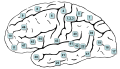
Brodmann area 4 (orange)
Ba4.png
Brodmann area 4 of human brain.
Details
Part of Precentral gyrus
Identifiers
Latin area gigantopyramidalis
NeuroNames 2394, 1014
NeuroLex ID birnlex_1735
FMA 68600
Anatomical terms of neuroanatomy

Brodmann area 4 refers to the primary motor cortex of the human brain. It is located in the posterior portion of the frontal lobe.

Contents

Brodmann area 4 is part of the precentral gyrus. The borders of this area are: the precentral sulcus in front (anteriorly), the medial longitudinal fissure at the top (medially), the central sulcus in back (posteriorly), and the lateral sulcus along the bottom (laterally).

This area of cortex, as shown by Wilder Penfield and others, has the pattern of a homunculus. That is, the legs and trunk fold over the midline; the arms and hands are along the middle of the area shown here; and the face is near the bottom of the figure. Because Brodmann area 4 is in the same general location as primary motor cortex, the homunculus here is called the motor homunculus.

The term area 4 of Brodmann-1909 refers to a cytoarchitecturally defined portion of the frontal lobe of the guenon. It is located predominantly in the precentral gyrus. Brodmann-1909 regarded it as topographically and cytoarchitecturally homologous to the human gigantopyramidal area 4 and noted that it occupies a much greater fraction of the frontal lobe in the monkey than in the human.

Distinctive features (Brodmann-1905): the cortex is unusually thick; the layers are not distinct; the cells are relatively sparsely distributed; giant pyramidal (Betz) cells are present in the internal pyramidal layer (V); lack of an internal granular layer (IV) such that the boundary between the external pyramidal layer (III) and the internal pyramidal layer (V) is indistinct; lack of a distinct external granular layer (II); a gradual transition from the multiform layer (VI) to the subcortical white matter. [1]

Image

See also

Related Research Articles

<span class="mw-page-title-main">Cingulate cortex</span> Part of the brain within the cerebral cortex

The cingulate cortex is a part of the brain situated in the medial aspect of the cerebral cortex. The cingulate cortex includes the entire cingulate gyrus, which lies immediately above the corpus callosum, and the continuation of this in the cingulate sulcus. The cingulate cortex is usually considered part of the limbic lobe.

<span class="mw-page-title-main">Brodmann area</span> Region of the brain

A Brodmann area is a region of the cerebral cortex, in the human or other primate brain, defined by its cytoarchitecture, or histological structure and organization of cells. The concept was first introduced by the German anatomist Korbinian Brodmann in the early 20th century. Brodmann mapped the human brain based on the varied cellular structure across the cortex and identified 52 distinct regions, which he numbered 1 to 52. These regions, or Brodmann areas, correspond with diverse functions including sensation, motor control, and cognition.

<span class="mw-page-title-main">Brodmann area 23</span>

Brodmann area 23 (BA23) is a region in the brain that lies inside the posterior cingulate cortex. It lies between Brodmann area 30 and Brodmann area 31 and is located on the medial wall of the cingulate gyrus between the callosal sulcus and the cingulate sulcus.

<span class="mw-page-title-main">Brodmann area 6</span>

Brodmann area 6 (BA6) is part of the frontal cortex in the human brain. Situated just anterior to the primary motor cortex (BA4), it is composed of the premotor cortex and, medially, the supplementary motor area (SMA). This large area of the frontal cortex is believed to play a role in planning complex, coordinated movements.

<span class="mw-page-title-main">Brodmann area 8</span> Brain area

Brodmann area 8 is one of Brodmann's cytologically defined regions of the brain. It is involved in planning complex movements.

<span class="mw-page-title-main">Brodmann area 10</span> Brain area

Brodmann area 10 is the anterior-most portion of the prefrontal cortex in the human brain. BA10 was originally defined broadly in terms of its cytoarchitectonic traits as they were observed in the brains of cadavers, but because modern functional imaging cannot precisely identify these boundaries, the terms anterior prefrontal cortex, rostral prefrontal cortex and frontopolar prefrontal cortex are used to refer to the area in the most anterior part of the frontal cortex that approximately covers BA10—simply to emphasize the fact that BA10 does not include all parts of the prefrontal cortex.

<span class="mw-page-title-main">Brodmann area 44</span> Brain area

Brodmann area 44, or BA44, is part of the frontal cortex in the human brain. Situated just anterior to premotor cortex (BA6) and on the lateral surface, inferior to BA9.

<span class="mw-page-title-main">Brodmann area 5</span> Brain area

Brodmann area 5 is one of Brodmann's cytoarchitectural defined regions of the brain. It is involved in somatosensory processing, movement and association, and is part of the posterior parietal cortex.

<span class="mw-page-title-main">Brodmann area 7</span> Brain area

Brodmann area 7 is one of Brodmann's cytologically defined regions of the brain corresponding to precuneus and superior parietal lobule (SPL). It is involved in locating objects in space. It serves as a point of convergence between vision and proprioception to determine where objects are in relation to parts of the body.

<span class="mw-page-title-main">Brodmann area 19</span>

Brodmann area 19, or BA 19, is part of the occipital lobe cortex in the human brain. Along with area 18, it comprises the extrastriate cortex. In humans with normal sight, extrastriate cortex is a visual association area, with feature-extracting, shape recognition, attentional, and multimodal integrating functions.

<span class="mw-page-title-main">Brodmann area 20</span>

Brodmann area 20, or BA20, is part of the temporal cortex in the human brain. The region encompasses most of the ventral temporal cortex, a region believed to play a part in high-level visual processing and recognition memory.

<span class="mw-page-title-main">Brodmann area 11</span> Brain area

Brodmann area 11 is one of Brodmann's cytologically defined regions of the brain. It is in the orbitofrontal cortex which is above the eye sockets (orbitae). It is involved in decision making, processing rewards, and encoding new information into long-term memory.

<span class="mw-page-title-main">Inferior frontal gyrus</span> Part of the brains prefrontal cortex

The inferior frontal gyrus (IFG),, is the lowest positioned gyrus of the frontal gyri, of the frontal lobe, and is part of the prefrontal cortex.

<span class="mw-page-title-main">Precentral gyrus</span> Motor gyrus of the posterior frontal lobe of the brain

The precentral gyrus is a prominent gyrus on the surface of the posterior frontal lobe of the brain. It is the site of the primary motor cortex that in humans is cytoarchitecturally defined as Brodmann area 4.

<span class="mw-page-title-main">Lobes of the brain</span> Parts of the cerebrum

The lobes of the brain are the major identifiable zones of the human cerebral cortex, and they comprise the surface of each hemisphere of the cerebrum. The two hemispheres are roughly symmetrical in structure, and are connected by the corpus callosum. They traditionally have been divided into four lobes, but are today considered as having six lobes each. The lobes are large areas that are anatomically distinguishable, and are also functionally distinct to some degree. Each lobe of the brain has numerous ridges, or gyri, and furrows, the sulci that constitute further subzones of the cortex. The expression "lobes of the brain" usually refers only to those of the cerebrum, not to the distinct areas of the cerebellum.

<span class="mw-page-title-main">Brodmann area 24</span> Brain area

Brodmann area 24 is part of the anterior cingulate in the human brain.

<span class="mw-page-title-main">Brodmann area 32</span> Brain area

The Brodmann area 32, also known in the human brain as the dorsal anterior cingulate area 32, refers to a subdivision of the cytoarchitecturally defined cingulate cortex. In the human it forms an outer arc around the anterior cingulate gyrus. The cingulate sulcus defines approximately its inner boundary and the superior rostral sulcus (H) its ventral boundary; rostrally it extends almost to the margin of the frontal lobe. Cytoarchitecturally it is bounded internally by the ventral anterior cingulate area 24, externally by medial margins of the agranular frontal area 6, intermediate frontal area 8, granular frontal area 9, frontopolar area 10, and prefrontal area 11-1909. (Brodmann19-09).

<span class="mw-page-title-main">Brodmann area 12</span> Brain area

Brodmann area 12 is a subdivision of the cerebral cortex of the guenon defined on the basis of cytoarchitecture. It occupies the most rostral portion of the frontal lobe. Brodmann-1909 did not regard it as homologous, either topographically or cytoarchitecturally, to rostral area 12 of the human. Distinctive features (Brodmann-1905): a quite distinct internal granular layer (IV) separates slender pyramidal cells of the external pyramidal layer (III) and the internal pyramidal layer (V); the multiform layer (VI) is expanded, contains widely dispersed spindle cells and merges gradually with the underlying cortical white matter; all cells, including the pyramidal cells of the external and internal pyramidal layers are inordinately small; the internal pyramidal layer (V) also contains spindle cells in groups of two to five located close to its border with the internal granular layer (IV).

References

  1. Korbinian Brodmann (1909). Vergleichende Lokalisationslehre der Grosshirnrinde in ihren Prinzipien dargestellt auf Grund des Zellenbaues. Leipzig: Barth. OCLC   14796098. (English translation available by Laurence Garey as Brodmann's 'Localisation in the cerebral cortex' (1994) OCLC   39219080 and later editions)