Uncus

Last updated
Uncus
Gray727 uncus of parahippocampal gyrus.png
Medial surface of left cerebral hemisphere. Uncus is shown in orange.
Human brain inferior-medial view description 3.JPG
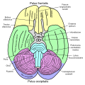
Human brain inferior-medial view (Uncus is #5)
Identifiers
NeuroNames 40
TA98 A14.1.09.235
TA2 5516
FMA 74884
Anatomical terms of neuroanatomy

The uncus is an anterior extremity of the parahippocampal gyrus. It is separated from the apex of the temporal lobe by a slight fissure called the incisura temporalis (also called rhinal sulcus [1] ).

Contents

Although superficially continuous with the hippocampal gyrus, the uncus forms morphologically a part of the rhinencephalon.

An important landmark that crosses the inferior surface of the uncus is the band of Giacomini. [2]

The term comes from the Latin word uncus, meaning hook, and it was coined by Félix Vicq-d'Azyr (1748–1794). [3]

Clinical significance

The part of the olfactory cortex that is on the temporal lobe covers the area of the uncus, which leads into the two significant clinical aspects of the uncus: uncinate fits and uncal herniations.

The landmark that helps you find the amygdala on a coronal section of the brain.

Function

A sparse amount of literature exists to propose a comprehensive overview of the functionality of the uncus. A study has indicated that psychotic-like experiences were associated with reduced expansion within the uncus between the ages of 14 and 19 in cannabis-using individuals. [4]

Additional images

Related Research Articles

Articles related to anatomy include:

<span class="mw-page-title-main">Brainstem</span> Posterior part of the brain, adjoining and structurally continuous

The brainstem is the stalk-like part of the brain that interconnects the cerebrum and diencephalon with the spinal cord. In the human brain, the brainstem is composed of the midbrain, the pons, and the medulla oblongata. The midbrain is continuous with the thalamus of the diencephalon through the tentorial notch.

<span class="mw-page-title-main">Temporal lobe</span> One of the four lobes of the mammalian brain

The temporal lobe is one of the four major lobes of the cerebral cortex in the brain of mammals. The temporal lobe is located beneath the lateral fissure on both cerebral hemispheres of the mammalian brain.

<span class="mw-page-title-main">Midbrain</span> Forward-most portion of the brainstem

The midbrain or mesencephalon is the rostral-most portion of the brainstem connecting the diencephalon and cerebrum with the pons. It consists of the cerebral peduncles, tegmentum, and tectum.

<span class="mw-page-title-main">Rhinencephalon</span>

In animal anatomy, the rhinencephalon, also called the smell-brain or olfactory brain, is a part of the brain involved with smell. It forms the paleocortex and is rudimentary in the human brain.

<span class="mw-page-title-main">Internal capsule</span> White matter structure situated in the inferomedial part of each cerebral hemisphere of the brain

The internal capsule is a white matter structure situated in the inferomedial part of each cerebral hemisphere of the brain. It carries information past the basal ganglia, separating the caudate nucleus and the thalamus from the putamen and the globus pallidus. The internal capsule contains both ascending and descending axons, going to and coming from the cerebral cortex. It also separates the caudate nucleus and the putamen in the dorsal striatum, a brain region involved in motor and reward pathways.

<span class="mw-page-title-main">Limbic lobe</span> Region of a cerebral cortex

The limbic lobe is an arc-shaped cortical region of the limbic system, on the medial surface of each cerebral hemisphere of the mammalian brain, consisting of parts of the frontal, parietal and temporal lobes. The term is ambiguous, with some authors including the paraterminal gyrus, the subcallosal area, the cingulate gyrus, the parahippocampal gyrus, the dentate gyrus, the hippocampus and the subiculum; while the Terminologia Anatomica includes the cingulate sulcus, the cingulate gyrus, the isthmus of cingulate gyrus, the fasciolar gyrus, the parahippocampal gyrus, the parahippocampal sulcus, the dentate gyrus, the fimbrodentate sulcus, the fimbria of hippocampus, the collateral sulcus, and the rhinal sulcus, and omits the hippocampus.

<span class="mw-page-title-main">Corticobulbar tract</span> Motor pathway in the brain connecting the motor cortex to the medullary pyramids

In neuroanatomy, the corticobulbartract is a two-neuron white matter motor pathway connecting the motor cortex in the cerebral cortex to the medullary pyramids, which are part of the brainstem's medulla oblongata region, and are primarily involved in carrying the motor function of the non-oculomotor cranial nerves. The corticobulbar tract is one of the pyramidal tracts, the other being the corticospinal tract.

<span class="mw-page-title-main">Precentral gyrus</span> Motor gyrus of the posterior frontal lobe of the brain

The precentral gyrus is a prominent gyrus on the surface of the posterior frontal lobe of the brain. It is the site of the primary motor cortex that in humans is cytoarchitecturally defined as Brodmann area 4.

<span class="mw-page-title-main">Cerebellar tentorium</span> Vertebrate brain structure separating the cerebellum from the occipital lobes

The cerebellar tentorium or tentorium cerebelli is an extension of the dura mater between the inferior aspect of the occipital lobes and the superior aspect of the cerebellum. The free border of the tentorium gives passage to the midbrain.

<span class="mw-page-title-main">Lobes of the brain</span> Parts of the cerebrum

The lobes of the brain are the major identifiable zones of the human cerebral cortex, and they comprise the surface of each hemisphere of the cerebrum. The two hemispheres are roughly symmetrical in structure, and are connected by the corpus callosum. They traditionally have been divided into four lobes, but are today considered as having six lobes each. The lobes are large areas that are anatomically distinguishable, and are also functionally distinct to some degree. Each lobe of the brain has numerous ridges, or gyri, and furrows, the sulci that constitute further subzones of the cortex. The expression "lobes of the brain" usually refers only to those of the cerebrum, not to the distinct areas of the cerebellum.

<span class="mw-page-title-main">Brain herniation</span> Potentially deadly side effect of very high pressure within the skull

Brain herniation is a potentially deadly side effect of very high pressure within the skull that occurs when a part of the brain is squeezed across structures within the skull. The brain can shift across such structures as the falx cerebri, the tentorium cerebelli, and even through the foramen magnum. Herniation can be caused by a number of factors that cause a mass effect and increase intracranial pressure (ICP): these include traumatic brain injury, intracranial hemorrhage, or brain tumor.

<span class="mw-page-title-main">Posterior cerebral artery</span> Artery which supplies blood to the occipital lobe of the brain

The posterior cerebral artery (PCA) is one of a pair of cerebral arteries that supply oxygenated blood to the occipital lobe, part of the back of the human brain. The two arteries originate from the distal end of the basilar artery, where it bifurcates into the left and right posterior cerebral arteries. These anastomose with the middle cerebral arteries and internal carotid arteries via the posterior communicating arteries.

<span class="mw-page-title-main">Duret haemorrhages</span> Medical condition

Duret haemorrhages are small linear areas of bleeding in the midbrain and upper pons of the brainstem. They are caused by a traumatic downward displacement of the brainstem.

<span class="mw-page-title-main">Rhinal sulcus</span>

In the human brain, the entorhinal cortex appears as a longitudinal elevation anterior to the parahippocampal gyrus, with a corresponding internal furrow, the external rhinal sulcus, separating it from the inferiolateral surface of the hemisphere close to the lamina terminalis. It is analogous to the collateral fissure found further caudally in the inferior part of the temporal lobe.

<span class="mw-page-title-main">Olfactory tract</span> Part of the olfactory system

The olfactory tract is a bilateral bundle of afferent nerve fibers from the mitral and tufted cells of the olfactory bulb that connects to several target regions in the brain, including the piriform cortex, amygdala, and entorhinal cortex. It is a narrow white band, triangular on coronal section, the apex being directed upward.

Kernohan's notch is a cerebral peduncle indentation associated with some forms of transtentorial herniation. It is a secondary condition caused by a primary injury on the opposite hemisphere of the brain. Kernohan's notch is an ipsilateral condition, in that a left-sided primary lesion evokes motor impairment in the left side of the body and a right-sided primary injury evokes motor impairment in the right side of the body. The seriousness of Kernohan's notch varies depending on the primary problem causing it, which may range from benign brain tumors to advanced subdural hematoma.

<span class="mw-page-title-main">Tentorial notch</span> Brain structure in humans and some mammals

The tentorial notch refers to the anterior opening between the free edge of the cerebellar tentorium and the clivus for the passage of the brainstem.

<span class="mw-page-title-main">Anatomical terms of neuroanatomy</span> Terminology used to describe the central and peripheral nervous systems

This article describes anatomical terminology that is used to describe the central and peripheral nervous systems - including the brain, brainstem, spinal cord, and nerves.

References

PD-icon.svgThis article incorporates text in the public domain from page 826 of the 20th edition of Gray's Anatomy (1918)

  1. Smith, Callum. "Rhinal sulcus | Radiology Reference Article | Radiopaedia.org". Radiopaedia. Retrieved 2022-08-15.
  2. Pfleger, René. "Uncus | Radiology Reference Article | Radiopaedia.org". Radiopaedia. Retrieved 17 October 2019.
  3. JC Tamraz, YG Comair. Atlas of Regional Anatomy of the Brain Using MRI (2006), p 8.
  4. Yu, Tao (2020). "Cannabis-associated psychotic-like experiences are mediated by developmental changes in the parahippocampal gyrus" (PDF). Journal of the American Academy of Child and Adolescent Psychiatry. 59 (5): 642–649. doi:10.1016/j.jaac.2019.05.034. PMID   31326579. S2CID   198135290 . Retrieved 15 August 2020.