Brodmann area 47

Last updated
Brodmann area 47
Brodmann Cytoarchitectonics 47.png
Brodmann area 47.png
Details
Identifiers
Latin area orbitalis
NeuroLex ID birnlex_1779
FMA 68644
Anatomical terms of neuroanatomy

Brodmann area 47, or BA47, is part of the frontal cortex in the human brain. It curves from the lateral surface of the frontal lobe into the ventral (orbital) frontal cortex. It is below areas BA10 and BA45, and beside BA11. This cytoarchitectonic region most closely corresponds to the gyral region the orbital part of inferior frontal gyrus, although these regions are not equivalent. Pars orbitalis is not based on cytoarchitectonic distinctions, and rather is defined according to gross anatomical landmarks. Despite a clear distinction, these two terms are often used liberally in peer-reviewed research journals.

Contents

BA47 is also known as orbital area 47. In the human, on the orbital surface it surrounds the caudal portion of the orbital sulcus (H) from which it extends laterally into the orbital part of inferior frontal gyrus (H). Cytoarchitectonically it is bounded caudally by the triangular area 45, medially by the prefrontal area 11 of Brodmann-1909, and rostrally by the frontopolar area 10 (Brodmann-1909).

It incorporates the region that Brodmann identified as "Area 12" in the monkey, and therefore, following the suggestion of Michael Petrides, some contemporary neuroscientists refer to the region as "BA47/12".

BA47 has been implicated in the processing of syntax in oral and sign languages, musical syntax, and semantic aspects of language.

Functions and Clinical Considerations

Language Processing and Comprehension

A major function of BA47 is language processing and comprehension. Although Broca’s and Wernicke’s areas are often the major foci of neuroanatomical studies related to language, research has discovered that these two areas are not as integral to language comprehension as originally thought; other structures like BA47 play a major role. [1] [2] Specifically, BA47 is active in tasks regarding semantics, or identifying the meaning of words and sentences. [3] [4] [5] To understand language semantics, consider Dapretto and Bookheimer’s (1999) study where participants needed to identify that there was a difference between the sentences, “The man was attacked by the Doberman,” and “The man was attacked by the Pitbull.” While sentence form was similar, the words Doberman and Pitbull had different meanings, specifically dog breeds. [6] This is indicative of a change in semantics. Having defined what semantics are, it is important to identify the functional limitations of individuals with damage to BA47. Patients with lesions to BA47 reported difficulty engaging in tasks that required one to process words as well as tasks that required one to be familiar with grammatical rules. [7]

Studies have determined that BA47 is involved in processing more than just spoken language. Considering BA47’s role in language semantics, it is essential to note that this function is not related only to oral communication; BA47 is also important for identifying semantics in sign language. [8] Specifically, BA47 plays a role in helping us determine what spoken words mean as well as what signed words mean. This finding that similar areas of the brain are active when processing different types of linguistic information is especially interesting considering the fact that the sensory modalities involved in spoken and sign language are different, the former involves audition and the latter involves vision. [9] Furthermore, in addition to language processing, BA47 helps us process music. Levitin and Menon (2003) found that BA47 showed greater activation when individuals were presented with “scrambled” sounds that violated their expectations versus sounds that went together and confirmed their expectations. [10] That is, with disrupted musical structure, participants required more brain processing for musical comprehension, and that happened in BA47.

Emotional Recognition

BA47 is thought to be related to the recognition of emotions. The emotions thought to be recognized by BA47 are fear, disgust, and anger. This hypothesis was tested be using an fMRI experiment on six healthy individuals. [11] They were shown eight faces showing the emotions of fear, disgust, anger, or neutral. The fMRI image was taken after the subjects were shown the face for 3 seconds. BA47 showed increased activity when the subject was shown the emotions of fear, disgust, and anger.

See also

Further reading

Related Research Articles

<span class="mw-page-title-main">Aphasia</span> Inability to comprehend or formulate language

In aphasia, a person may be unable to comprehend or unable to formulate language because of damage to specific brain regions. The major causes are stroke and head trauma; prevalence is hard to determine but aphasia due to stroke is estimated to be 0.1–0.4% in the Global North. Aphasia can also be the result of brain tumors, epilepsy, autoimmune neurological diseases, brain infections, or neurodegenerative diseases.

<span class="mw-page-title-main">Language center</span> Speech processing areas of the brain

In neuroscience and psychology, the term language center refers collectively to the areas of the brain which serve a particular function for speech processing and production. Language is a core system that gives humans the capacity to solve difficult problems and provides them with a unique type of social interaction. Language allows individuals to attribute symbols to specific concepts, and utilize them through sentences and phrases that follow proper grammatical rules. Finally, speech is the mechanism by which language is orally expressed.

<span class="mw-page-title-main">Broca's area</span> Speech production region in the dominant hemisphere of the hominid brain

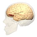
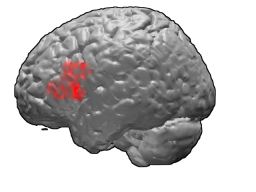
Broca's area, or the Broca area, is a region in the frontal lobe of the dominant hemisphere, usually the left, of the brain with functions linked to speech production.

<span class="mw-page-title-main">Neurolinguistics</span> Neuroscience and linguistics-related studies

Neurolinguistics is the study of neural mechanisms in the human brain that control the comprehension, production, and acquisition of language. As an interdisciplinary field, neurolinguistics draws methods and theories from fields such as neuroscience, linguistics, cognitive science, communication disorders and neuropsychology. Researchers are drawn to the field from a variety of backgrounds, bringing along a variety of experimental techniques as well as widely varying theoretical perspectives. Much work in neurolinguistics is informed by models in psycholinguistics and theoretical linguistics, and is focused on investigating how the brain can implement the processes that theoretical and psycholinguistics propose are necessary in producing and comprehending language. Neurolinguists study the physiological mechanisms by which the brain processes information related to language, and evaluate linguistic and psycholinguistic theories, using aphasiology, brain imaging, electrophysiology, and computer modeling.

Aphasiology is the study of language impairment usually resulting from brain damage, due to neurovascular accident—hemorrhage, stroke—or associated with a variety of neurodegenerative diseases, including different types of dementia. These specific language deficits, termed aphasias, may be defined as impairments of language production or comprehension that cannot be attributed to trivial causes such as deafness or oral paralysis. A number of aphasias have been described, but two are best known: expressive aphasia and receptive aphasia.

<span class="mw-page-title-main">Cingulate cortex</span> Part of the brain within the cerebral cortex

The cingulate cortex is a part of the brain situated in the medial aspect of the cerebral cortex. The cingulate cortex includes the entire cingulate gyrus, which lies immediately above the corpus callosum, and the continuation of this in the cingulate sulcus. The cingulate cortex is usually considered part of the limbic lobe.

<span class="mw-page-title-main">Brodmann area 10</span> Brain area

Brodmann area 10 is the anterior-most portion of the prefrontal cortex in the human brain. BA10 was originally defined broadly in terms of its cytoarchitectonic traits as they were observed in the brains of cadavers, but because modern functional imaging cannot precisely identify these boundaries, the terms anterior prefrontal cortex, rostral prefrontal cortex and frontopolar prefrontal cortex are used to refer to the area in the most anterior part of the frontal cortex that approximately covers BA10—simply to emphasize the fact that BA10 does not include all parts of the prefrontal cortex.

<span class="mw-page-title-main">Brodmann area 44</span> Brain area

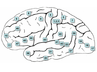
Brodmann area 44, or BA44, is part of the frontal cortex in the human brain. Situated just anterior to premotor cortex (BA6) and on the lateral surface, inferior to BA9.

<span class="mw-page-title-main">Brodmann area 45</span> Brain area

Brodmann area 45 (BA45), is part of the frontal cortex in the human brain. It is situated on the lateral surface, inferior to BA9 and adjacent to BA46.

<span class="mw-page-title-main">Brodmann area 46</span> Brain area

Brodmann area 46, or BA46, is part of the frontal cortex in the human brain. It is between BA10 and BA45.

<span class="mw-page-title-main">Wernicke's area</span> Speech comprehension region in the dominant hemisphere of the hominid brain

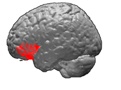
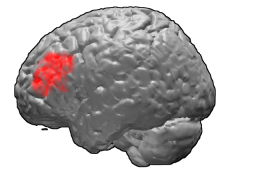
Wernicke's area, also called Wernicke's speech area, is one of the two parts of the cerebral cortex that are linked to speech, the other being Broca's area. It is involved in the comprehension of written and spoken language, in contrast to Broca's area, which is primarily involved in the production of language. It is traditionally thought to reside in Brodmann area 22, which is located in the superior temporal gyrus in the dominant cerebral hemisphere, which is the left hemisphere in about 95% of right-handed individuals and 70% of left-handed individuals.

<span class="mw-page-title-main">Inferior frontal gyrus</span> Part of the brains prefrontal cortex

The inferior frontal gyrus (IFG),, is the lowest positioned gyrus of the frontal gyri, of the frontal lobe, and is part of the prefrontal cortex.

<span class="mw-page-title-main">Arcuate fasciculus</span> Neural pathway connecting Brocas area and Wernickes area

In neuroanatomy, the arcuate fasciculus is a bundle of axons that generally connects the Broca's area and the Wernicke's area in the brain. It is an association fiber tract connecting caudal temporal cortex and inferior frontal lobe.

<span class="mw-page-title-main">Language processing in the brain</span> How humans use words to communicate

In psycholinguistics, language processing refers to the way humans use words to communicate ideas and feelings, and how such communications are processed and understood. Language processing is considered to be a uniquely human ability that is not produced with the same grammatical understanding or systematicity in even human's closest primate relatives.

The language module or language faculty is a hypothetical structure in the human brain which is thought to contain innate capacities for language, originally posited by Noam Chomsky. There is ongoing research into brain modularity in the fields of cognitive science and neuroscience, although the current idea is much weaker than what was proposed by Chomsky and Jerry Fodor in the 1980s. In today's terminology, 'modularity' refers to specialisation: language processing is specialised in the brain to the extent that it occurs partially in different areas than other types of information processing such as visual input. The current view is, then, that language is neither compartmentalised nor based on general principles of processing. It is modular to the extent that it constitutes a specific cognitive skill or area in cognition.

<span class="mw-page-title-main">Prefrontal cortex</span> Part of the brain responsible for personality, decision-making, and social behavior

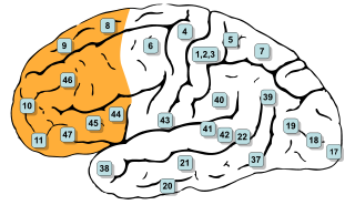
In mammalian brain anatomy, the prefrontal cortex (PFC) covers the front part of the frontal lobe of the cerebral cortex. It is the association cortex in the frontal lobe. The PFC contains the Brodmann areas BA8, BA9, BA10, BA11, BA12, BA13, BA14, BA24, BA25, BA32, BA44, BA45, BA46, and BA47.

<span class="mw-page-title-main">Brodmann area 22</span>

Brodmann area 22 is a Brodmann's area that is cytoarchitecturally located in the posterior superior temporal gyrus of the brain. In the left cerebral hemisphere, it is one portion of Wernicke's area. The left hemisphere BA22 helps with generation and understanding of individual words. On the right side of the brain, BA22 helps to discriminate pitch and sound intensity, both of which are necessary to perceive melody and prosody. Wernicke's area is active in processing language and consists of the left Brodmann area 22 and Brodmann area 40, the supramarginal gyrus.

Neuroscience of multilingualism is the study of multilingualism within the field of neurology. These studies include the representation of different language systems in the brain, the effects of multilingualism on the brain's structural plasticity, aphasia in multilingual individuals, and bimodal bilinguals. Neurological studies of multilingualism are carried out with functional neuroimaging, electrophysiology, and through observation of people who have suffered brain damage.

<span class="mw-page-title-main">Orbital part of inferior frontal gyrus</span>

The orbital part of inferior frontal gyrus also known as the pars orbitalis is the orbital part of the inferior frontal gyrus.

Nina Dronkers is an American cognitive neuroscientist. She is known for her studies of aphasia and their application for understanding brain systems involved in normal language abilities. She is a professor in the Psychology Department at the University of California, Berkeley, and a faculty member of the Helen Wills Neuroscience Institute. She is also an Emerita Research Career Scientist of the Veterans Administration Northern California Health Care System where she established the Center for Aphasia and Related Disorders. She serves as a consultant for the Memory and Aging Center at UCSF Medical Center. In addition, she is an adjunct professor in the Department of Neurology, University of California, Davis, School of Medicine. She has published over 120 scientific papers and is the co-author with Lise Menn of a textbook: Psycholinguistics: Introduction and Applications, Second edition.

References

  1. Dronkers, Nina F; Wilkins, David P; Van Valin, Robert D; Redfern, Brenda B; Jaeger, Jeri J (May 2004). "Lesion analysis of the brain areas involved in language comprehension". Cognition. 92 (1–2): 145–177. doi:10.1016/j.cognition.2003.11.002. hdl: 11858/00-001M-0000-0012-6912-A . PMID   15037129. S2CID   10919645.
  2. Ardila, Alfredo; Bernal, Byron; Rosselli, Monica (February 2017). "Should Broca's area include Brodmann area 47?". Psicothema. 29 (1): 73–77. doi:10.7334/psicothema2016.11. PMID   28126062.
  3. Dapretto, Mirella; Bookheimer, Susan Y (October 1999). "Form and Content". Neuron. 24 (2): 427–432. doi: 10.1016/S0896-6273(00)80855-7 . PMID   10571235. S2CID   18742085.
  4. Dronkers, Nina F; Wilkins, David P; Van Valin, Robert D; Redfern, Brenda B; Jaeger, Jeri J (May 2004). "Lesion analysis of the brain areas involved in language comprehension". Cognition. 92 (1–2): 145–177. doi:10.1016/j.cognition.2003.11.002. hdl: 11858/00-001M-0000-0012-6912-A . PMID   15037129. S2CID   10919645.
  5. Ardila, Alfredo; Bernal, Byron; Rosselli, Monica (February 2017). "Should Broca's area include Brodmann area 47?". Psicothema. 29 (1): 73–77. doi:10.7334/psicothema2016.11. PMID   28126062.
  6. Dapretto, Mirella; Bookheimer, Susan Y (October 1999). "Form and Content". Neuron. 24 (2): 427–432. doi: 10.1016/S0896-6273(00)80855-7 . PMID   10571235. S2CID   18742085.
  7. Dronkers, Nina F; Wilkins, David P; Van Valin, Robert D; Redfern, Brenda B; Jaeger, Jeri J (May 2004). "Lesion analysis of the brain areas involved in language comprehension". Cognition. 92 (1–2): 145–177. doi:10.1016/j.cognition.2003.11.002. hdl: 11858/00-001M-0000-0012-6912-A . PMID   15037129. S2CID   10919645.
  8. Petitto, L. A.; Zatorre, R. J.; Gauna, K.; Nikelski, E. J.; Dostie, D.; Evans, A. C. (2000-12-05). "Speech-like cerebral activity in profoundly deaf people processing signed languages: Implications for the neural basis of human language". Proceedings of the National Academy of Sciences. 97 (25): 13961–13966. doi: 10.1073/pnas.97.25.13961 . ISSN   0027-8424. PMC   17683 . PMID   11106400.
  9. Petitto, L. A.; Zatorre, R. J.; Gauna, K.; Nikelski, E. J.; Dostie, D.; Evans, A. C. (2000-12-05). "Speech-like cerebral activity in profoundly deaf people processing signed languages: Implications for the neural basis of human language". Proceedings of the National Academy of Sciences. 97 (25): 13961–13966. doi: 10.1073/pnas.97.25.13961 . ISSN   0027-8424. PMC   17683 . PMID   11106400.
  10. Levitin, D (December 2003). "Musical structure is processed in "language" areas of the brain: a possible role for Brodmann Area 47 in temporal coherence". NeuroImage. 20 (4): 2142–2152. doi:10.1016/j.neuroimage.2003.08.016. PMID   14683718. S2CID   14691229.
  11. Sprengelmeyer, R.; Rausch, M.; Eysel, U. T.; Przuntek, H. (1998-10-22). "Neural structures associated with recognition of facial expressions of basic emotions". Proceedings of the Royal Society of London. Series B: Biological Sciences. 265 (1409): 1927–1931. doi:10.1098/rspb.1998.0522. ISSN   0962-8452. PMC   1689486 . PMID   9821359.