Brodmann area 38

Last updated
Brodmann area 38
Brodmann area 38.png
Brodmann Cytoarchitectonics 38.png
Details
Identifiers
Latin area temporopolaris
NeuroLex ID birnlex_1771
FMA 68635
Anatomical terms of neuroanatomy

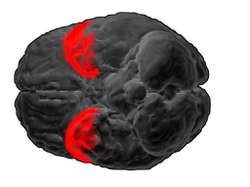
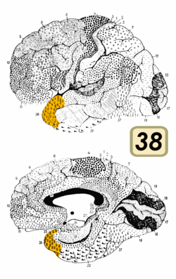
Brodmann area 38, also BA38 or temporopolar area 38 (H), is part of the temporal cortex in the human brain. BA 38 is at the anterior end of the temporal lobe, known as the temporal pole.

Contents

BA38 is a subdivision of the cytoarchitecturally defined temporal region of cerebral cortex. It is located primarily in the most rostral portions of the superior temporal gyrus and the middle temporal gyrus. Cytoarchitecturally it is bounded caudally by the inferior temporal area 20, the middle temporal area 21, the superior temporal area 22 and the ectorhinal area 36 (Brodmann-1909).

The temporal pole is a paralimbic region involved in high level semantic representation and socio-emotional processing. The uncinate fasciculus provides a direct bidirectional path to the orbitofrontal cortex, allowing mnemonic representations stored in the temporal pole to bias decision making in the frontal lobe. The temporal pole appears to be a convergence zone where concepts (also known as semantic memories) that are stored in the ventral anterior temporal lobe are imbued with emotional significance and personal meaning. [1] In addition, concepts of individual people, abstracted away from the perceptual representations, are stored in a “face patch” in the temporal pole. This face patch is found in both non-human primates and humans. [2] This relates to early work showing that damage to the temporal pole can cause an amnestic prosopagnosia in which recognition of familiar faces is lost. [2]

Bilateral damage to the temporal poles, though rare, can cause dramatic changes in personality. Klüver-Bucy syndrome involves damage to the greater temporal pole as well as the amygdala. In this disorder, people and animals demonstrate fearlessness, hypersexuality, and hyperorality. [1]

This area is among the earliest affected by Alzheimer's disease, frontotemporal dementia, frontotemporal lobar degeneration, and is commonly involved at the start of temporal lobe seizures. [3]

Cytoarchitectonic and chemoarchitectonic studies find that it contains at least seven subareas, one of which, "TG", is unique to humans. [3]

See also

Related Research Articles

<span class="mw-page-title-main">Broca's area</span> Speech production region in the dominant hemisphere of the hominid brain

Broca's area, or the Broca area, is a region in the frontal lobe of the dominant hemisphere, usually the left, of the brain with functions linked to speech production.

<span class="mw-page-title-main">Cingulate cortex</span> Part of the brain within the cerebral cortex

The cingulate cortex is a part of the brain situated in the medial aspect of the cerebral cortex. The cingulate cortex includes the entire cingulate gyrus, which lies immediately above the corpus callosum, and the continuation of this in the cingulate sulcus. The cingulate cortex is usually considered part of the limbic lobe.

<span class="mw-page-title-main">Brodmann area</span> Region of the brain

A Brodmann area is a region of the cerebral cortex, in the human or other primate brain, defined by its cytoarchitecture, or histological structure and organization of cells. The concept was first introduced by the German anatomist Korbinian Brodmann in the early 20th century. Brodmann mapped the human brain based on the varied cellular structure across the cortex and identified 52 distinct regions, which he numbered 1 to 52. These regions, or Brodmann areas, correspond with diverse functions including sensation, motor control, and cognition.

<span class="mw-page-title-main">Brodmann area 23</span>

Brodmann area 23 (BA23) is a region in the brain that lies inside the posterior cingulate cortex. It lies between Brodmann area 30 and Brodmann area 31 and is located on the medial wall of the cingulate gyrus between the callosal sulcus and the cingulate sulcus.

<span class="mw-page-title-main">Parietal lobe</span> Part of the brain responsible for sensory input and some language processing

The parietal lobe is one of the four major lobes of the cerebral cortex in the brain of mammals. The parietal lobe is positioned above the temporal lobe and behind the frontal lobe and central sulcus.

<span class="mw-page-title-main">Temporal lobe</span> One of the four lobes of the mammalian brain

The temporal lobe is one of the four major lobes of the cerebral cortex in the brain of mammals. The temporal lobe is located beneath the lateral fissure on both cerebral hemispheres of the mammalian brain.

<span class="mw-page-title-main">Brodmann area 10</span> Brain area

Brodmann area 10 is the anterior-most portion of the prefrontal cortex in the human brain. BA10 was originally defined broadly in terms of its cytoarchitectonic traits as they were observed in the brains of cadavers, but because modern functional imaging cannot precisely identify these boundaries, the terms anterior prefrontal cortex, rostral prefrontal cortex and frontopolar prefrontal cortex are used to refer to the area in the most anterior part of the frontal cortex that approximately covers BA10—simply to emphasize the fact that BA10 does not include all parts of the prefrontal cortex.

<span class="mw-page-title-main">Brodmann area 44</span> Brain area

Brodmann area 44, or BA44, is part of the frontal cortex in the human brain. Situated just anterior to premotor cortex (BA6) and on the lateral surface, inferior to BA9.

<span class="mw-page-title-main">Brodmann area 19</span>

Brodmann area 19, or BA 19, is part of the occipital lobe cortex in the human brain. Along with area 18, it comprises the extrastriate cortex. In humans with normal sight, extrastriate cortex is a visual association area, with feature-extracting, shape recognition, attentional, and multimodal integrating functions.

<span class="mw-page-title-main">Brodmann area 21</span> Area of the temporal cortex

Brodmann area 21, or BA21, is part of the temporal cortex in the human brain. The region encompasses most of the lateral temporal cortex and is also known as middle temporal area 21. In the human it corresponds approximately to the middle temporal gyrus.

<span class="mw-page-title-main">Brodmann area 37</span>

Brodmann area 37, or BA37, is part of the temporal cortex in the human brain. It contains the fusiform gyrus which in turn contains the fusiform face area, an area important for the recognition of faces.

<span class="mw-page-title-main">Brodmann area 40</span> Part of the parietal cortex in the human brain

Brodmann area 40 (BA40) is part of the parietal cortex in the human brain. The inferior part of BA40 is in the area of the supramarginal gyrus, which lies at the posterior end of the lateral fissure, in the inferior lateral part of the parietal lobe.

<span class="mw-page-title-main">Inferior frontal gyrus</span> Part of the brains prefrontal cortex

The inferior frontal gyrus (IFG),, is the lowest positioned gyrus of the frontal gyri, of the frontal lobe, and is part of the prefrontal cortex.

<span class="mw-page-title-main">Brodmann area 4</span>

Brodmann area 4 refers to the primary motor cortex of the human brain. It is located in the posterior portion of the frontal lobe.

<span class="mw-page-title-main">Brodmann area 22</span>

Brodmann area 22 is a Brodmann's area that is cytoarchitecturally located in the posterior superior temporal gyrus of the brain. In the left cerebral hemisphere, it is one portion of Wernicke's area. The left hemisphere BA22 helps with generation and understanding of individual words. On the right side of the brain, BA22 helps to discriminate pitch and sound intensity, both of which are necessary to perceive melody and prosody. Wernicke's area is active in processing language and consists of the left Brodmann area 22 and Brodmann area 40, the supramarginal gyrus.

<span class="mw-page-title-main">Brodmann areas 35 and 36</span>

Brodmann area 35, together with Brodmann area 36, comprise the perirhinal cortex. They are cytoarchitecturally defined temporal regions of the cerebral cortex.

<span class="mw-page-title-main">Posterior cingulate cortex</span> Caudal part of the cingulate cortex of the brain

The posterior cingulate cortex (PCC) is the caudal part of the cingulate cortex, located posterior to the anterior cingulate cortex. This is the upper part of the "limbic lobe". The cingulate cortex is made up of an area around the midline of the brain. Surrounding areas include the retrosplenial cortex and the precuneus.

<span class="mw-page-title-main">Uncinate fasciculus</span> White matter tract in the human brain

The uncinate fasciculus is a white matter association tract in the human brain that connects parts of the limbic system such as the temporal pole, anterior parahippocampus, and amygdala in the temporal lobe with inferior portions of the frontal lobe such as the orbitofrontal cortex. Its function is unknown though it is affected in several psychiatric conditions. It is one of the last white matter tracts to mature in the human brain.

<span class="mw-page-title-main">Auditosensory cortex</span>

The auditosensory cortex is the part of the auditory system that is associated with the sense of hearing in humans. It occupies the bilateral primary auditory cortex in the temporal lobe of the mammalian brain. The term is used to describe Brodmann areas 41 and 42 together with the transverse temporal gyrus. The auditosensory cortex takes part in the reception and processing of auditory nerve impulses, which passes sound information from the thalamus to the brain. Abnormalities in this region are responsible for many disorders in auditory abilities, such as congenital deafness, true cortical deafness, primary progressive aphasia and auditory hallucination.

References

  1. 1 2 Olson, IR; Plotzker, A; Ezzyat, Y (2007). "The enigmatic temporal poles: A review of findings on social and emotional processing". Brain. 130 (7): 1718–1731. doi: 10.1093/brain/awm052 . PMID   17392317.
  2. 1 2 Von der Heide, RJ; Skipper, LM; Olson, IR (2013). "Anterior temporal face patches: A meta-analysis and empirical study". Frontiers in Human Neuroscience. 7: 17. doi: 10.3389/fnhum.2013.00017 . PMC   3561664 . PMID   23378834.
  3. 1 2 Ding, S. L.; Van Hoesen, G. W.; Cassell, M. D.; Poremba, A. (2009). "Parcellation of human temporal polar cortex: A combined analysis of multiple cytoarchitectonic, chemoarchitectonic, and pathological markers". The Journal of Comparative Neurology. 514 (6): 595–623. doi:10.1002/cne.22053. PMC   3665344 . PMID   19363802.