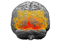
The visual cortex of the brain is the area of the cerebral cortex that processes visual information. It is located in the occipital lobe. Sensory input originating from the eyes travels through the lateral geniculate nucleus in the thalamus and then reaches the visual cortex. The area of the visual cortex that receives the sensory input from the lateral geniculate nucleus is the primary visual cortex, also known as visual area 1 (V1), Brodmann area 17, or the striate cortex. The extrastriate areas consist of visual areas 2, 3, 4, and 5.

A saccade is a quick, simultaneous movement of both eyes between two or more phases of focal points in the same direction. In contrast, in smooth-pursuit movements, the eyes move smoothly instead of in jumps; it could be associated with a shift in frequency of an emitted signal or a movement of a body part or device. Controlled cortically by the frontal eye fields (FEF), or subcortically by the superior colliculus, saccades serve as a mechanism for focal points, rapid eye movement, and the fast phase of optokinetic nystagmus. The word appears to have been coined in the 1880s by French ophthalmologist Émile Javal, who used a mirror on one side of a page to observe eye movement in silent reading, and found that it involves a succession of discontinuous individual movements.
Saccadic masking, also known as (visual) saccadic suppression, is the phenomenon in visual perception where the brain selectively blocks visual processing during eye movements in such a way that neither the motion of the eye nor the gap in visual perception is noticeable to the viewer.

The parietal lobe is one of the four major lobes of the cerebral cortex in the brain of mammals. The parietal lobe is positioned above the temporal lobe and behind the frontal lobe and central sulcus.

The auditory system is the sensory system for the sense of hearing. It includes both the sensory organs and the auditory parts of the sensory system.

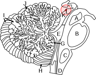
In neuroanatomy, the superior colliculus is a structure lying on the roof of the mammalian midbrain. In non-mammalian vertebrates, the homologous structure is known as the optic tectum or optic lobe. The adjective form tectal is commonly used for both structures.
Microsaccades are a kind of fixational eye movement. They are small, jerk-like, involuntary eye movements, similar to miniature versions of voluntary saccades. They typically occur during prolonged visual fixation, not only in humans, but also in animals with foveal vision. Microsaccade amplitudes vary from 2 to 120 arcminutes. The first empirical evidence for their existence was provided by Robert Darwin, the father of Charles Darwin.

In the scientific study of vision, smooth pursuit describes a type of eye movement in which the eyes remain fixated on a moving object. It is one of two ways that visual animals can voluntarily shift gaze, the other being saccadic eye movements. Pursuit differs from the vestibulo-ocular reflex, which only occurs during movements of the head and serves to stabilize gaze on a stationary object. Most people are unable to initiate pursuit without a moving visual signal. The pursuit of targets moving with velocities of greater than 30°/s tends to require catch-up saccades. Smooth pursuit is asymmetric: most humans and primates tend to be better at horizontal than vertical smooth pursuit, as defined by their ability to pursue smoothly without making catch-up saccades. Most humans are also better at downward than upward pursuit. Pursuit is modified by ongoing visual feedback.

The paramedian pontine reticular formation (PPRF) is a subset of neurons of the oral and caudal pontine reticular nuclei. With the abducens nucleus it makes up the horizontal gaze centre. It is situated in the pons adjacent to the abducens nucleus. It projects to the ipsilateral abducens nucleus, and contralateral oculomotor nucleus to mediate conjugate horizontal gaze and saccades.

Supplementary eye field (SEF) is the name for the anatomical area of the dorsal medial frontal lobe of the primate cerebral cortex that is indirectly involved in the control of saccadic eye movements. Evidence for a supplementary eye field was first shown by Schlag, and Schlag-Rey. Current research strives to explore the SEF's contribution to visual search and its role in visual salience. The SEF constitutes together with the frontal eye fields (FEF), the intraparietal sulcus (IPS), and the superior colliculus (SC) one of the most important brain areas involved in the generation and control of eye movements, particularly in the direction contralateral to their location. Its precise function is not yet fully known. Neural recordings in the SEF show signals related to both vision and saccades somewhat like the frontal eye fields and superior colliculus, but currently most investigators think that the SEF has a special role in high level aspects of saccade control, like complex spatial transformations, learned transformations, and executive cognitive functions.

The intraparietal sulcus (IPS) is located on the lateral surface of the parietal lobe, and consists of an oblique and a horizontal portion. The IPS contains a series of functionally distinct subregions that have been intensively investigated using both single cell neurophysiology in primates and human functional neuroimaging. Its principal functions are related to perceptual-motor coordination and visual attention, which allows for visually-guided pointing, grasping, and object manipulation that can produce a desired effect.
Attentional shift occurs when directing attention to a point increases the efficiency of processing of that point and includes inhibition to decrease attentional resources to unwanted or irrelevant inputs. Shifting of attention is needed to allocate attentional resources to more efficiently process information from a stimulus. Research has shown that when an object or area is attended, processing operates more efficiently. Task switching costs occur when performance on a task suffers due to the increased effort added in shifting attention. There are competing theories that attempt to explain why and how attention is shifted as well as how attention is moved through space in attentional control.

The medial dorsal nucleus is a large nucleus in the thalamus. It is separated from the other thalamic nuclei by the internal medullary lamina.

The posterior parietal cortex plays an important role in planned movements, spatial reasoning, and attention.
Auditory spatial attention is a specific form of attention, involving the focusing of auditory perception to a location in space.
Transsaccadic memory is the neural process that allows humans to perceive their surroundings as a seamless, unified image despite rapid changes in fixation points. Transsaccadic memory is a relatively new topic of interest in the field of psychology. Conflicting views and theories have spurred several types of experiments intended to explain transsaccadic memory and the neural mechanisms involved.
The anti-saccade (AS) task is a way of measuring how well the frontal lobe of the brain can control the reflexive saccade, or eye movement. Saccadic eye movement is primarily controlled by the frontal cortex.

Peter H. Schiller was a German-born neuroscientist. At the time of his death, he was a professor emeritus of Neuroscience in the Department of Brain and Cognitive Sciences at the Massachusetts Institute of Technology (MIT). Schiller is well known for his work on the behavioral, neurophysiological and pharmacological studies of the primate visual and oculomotor systems.
Michael E. Goldberg, also known as Mickey Goldberg, is an American neuroscientist and David Mahoney Professor at Columbia University. He is known for his work on the mechanisms of the mammalian eye in relation to brain activity. He served as president of the Society for Neuroscience from 2009 to 2010.
The corollary discharge theory (CD) of motion perception helps understand how the brain can detect motion through the visual system, even though the body is not moving. When a signal is sent from the motor cortex of the brain to the eye muscles, a copy of that signal is sent through the brain as well. The brain does this in order to distinguish real movements in the visual world from our own body and eye movement. The original signal and copy signal are then believed to be compared somewhere in the brain. Such a structure has not yet been identified, but it is believed to be the Medial Superior Temporal Area (MST). The original signal and copy need to be compared in order to determine if the change in vision was caused by eye movement or movement in the world. If the two signals cancel then no motion is perceived, but if they do not cancel then the residual signal is perceived as motion in the real world. Without a corollary discharge signal, the world would seem to spin around every time the eyes moved. It is important to note that corollary discharge and efference copy are sometimes used synonymously, they were originally coined for much different applications, with corollary discharge being used in a much broader sense.


