Superior temporal gyrus

Last updated
Superior temporal gyrus
Superior temporal gyrus.png
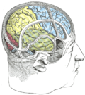
Superior temporal gyrus of the human brain.
Slide2HAN.JPG
Cerebrum. Lateral view. Deep dissection. Superior temporal gyrus is visible at the center.
Details
Part of Temporal lobe
Location Temporal lobe of the human brain
Artery middle cerebral
Identifiers
Latin gyrus temporalis superior
NeuroNames 136
NeuroLex ID birnlex_1648
TA98 A14.1.09.138
TA2 5489
FMA 61905
Anatomical terms of neuroanatomy

The superior temporal gyrus (STG [1] ) is one of three (sometimes two) gyri in the temporal lobe of the human brain, which is located laterally to the head, situated somewhat above the external ear.

Contents

The superior temporal gyrus is bounded by:

The superior temporal gyrus contains several important structures of the brain, including:

The superior temporal gyrus contains the auditory cortex, which is responsible for processing sounds. Specific sound frequencies map precisely onto the auditory cortex. This auditory (or tonotopic) map is similar to the homunculus map of the primary motor cortex. Some areas of the superior temporal gyrus are specialized for processing combinations of frequencies, and other areas are specialized for processing changes in amplitude or frequency. The superior temporal gyrus also includes Wernicke's area, which (in most people) is located in the left hemisphere. It is the major area involved in the comprehension of language. The superior temporal gyrus is involved in auditory processing, including language, but also has been implicated as a critical structure in social cognition. [2] [3]

Various parts of the STG might be referred to as anterior (aSTG), middle (mSTG), and posterior (pSTG).

Function

The superior temporal gyrus has been involved in the perception of emotions in facial stimuli. [2] [4] ) Furthermore, the superior temporal gyrus is an essential structure involved in auditory processing, as well as in the function of language in individuals who may have an impaired vocabulary, or are developing a sense of language. The superior temporal gyrus has been discovered to be an important structure in the pathway consisting of the amygdala and prefrontal cortex, which are all involved in social cognition processes. [5] [6] [7] Including the superior temporal gyrus, areas more anterior and dorsal within the temporal lobe have been linked to the ability of processing information the many changeable characteristics of a face. [8] Research conducted with the use of neuroimaging have found patients with schizophrenia have structural abnormalities in their superior temporal gyrus. [9]

fMRI analysis has evidenced a link between insight based problem solving and activity in the right anterior superior-temporal gyrus, specifically in relation to the sudden flash of understanding commonly referred to as an 'Aha!' moment. [10]

Social context

The superior temporal gyrus (STG) is important for language comprehension, but studies also suggest that it plays a functional role in the cocktail party effect. A magnetoencephalography study was conducted on participants that were exposed to five differing listening conditions each with a different level of background noise. It was discovered that the STG has a strong connection with the attended speech stream in a cocktail party setting. When the attended speech stream wasn’t disrupted by background noise a bilateral connection was displayed, but as more background noise was introduced the connection became left-hemisphere-dependent. [11]

Additional images

Related Research Articles

<span class="mw-page-title-main">Brodmann area</span> Region of the brain

A Brodmann area is a region of the cerebral cortex, in the human or other primate brain, defined by its cytoarchitecture, or histological structure and organization of cells. The concept was first introduced by the German anatomist Korbinian Brodmann in the early 20th century. Brodmann mapped the human brain based on the varied cellular structure across the cortex and identified 52 distinct regions, which he numbered 1 to 52. These regions, or Brodmann areas, correspond with diverse functions including sensation, motor control, and cognition.

<span class="mw-page-title-main">Temporal lobe</span> One of the four lobes of the mammalian brain

The temporal lobe is one of the four major lobes of the cerebral cortex in the brain of mammals. The temporal lobe is located beneath the lateral fissure on both cerebral hemispheres of the mammalian brain.

<span class="mw-page-title-main">Wernicke's area</span> Speech comprehension region in the dominant hemisphere of the hominid brain

Wernicke's area, also called Wernicke's speech area, is one of the two parts of the cerebral cortex that are linked to speech, the other being Broca's area. It is involved in the comprehension of written and spoken language, in contrast to Broca's area, which is primarily involved in the production of language. It is traditionally thought to reside in Brodmann area 22, which is located in the superior temporal gyrus in the dominant cerebral hemisphere, which is the left hemisphere in about 95% of right-handed individuals and 70% of left-handed individuals.

<span class="mw-page-title-main">Auditory system</span> Sensory system used for hearing

The auditory system is the sensory system for the sense of hearing. It includes both the sensory organs and the auditory parts of the sensory system.

<span class="mw-page-title-main">Fusiform gyrus</span> Gyrus of the temporal and occipital lobes of the brain

The fusiform gyrus, also known as the lateral occipitotemporal gyrus,is part of the temporal lobe and occipital lobe in Brodmann area 37. The fusiform gyrus is located between the lingual gyrus and parahippocampal gyrus above, and the inferior temporal gyrus below. Though the functionality of the fusiform gyrus is not fully understood, it has been linked with various neural pathways related to recognition. Additionally, it has been linked to various neurological phenomena such as synesthesia, dyslexia, and prosopagnosia.

<span class="mw-page-title-main">Inferior frontal gyrus</span> Part of the brains prefrontal cortex

The inferior frontal gyrus(IFG), (gyrus frontalis inferior), is the lowest positioned gyrus of the frontal gyri, of the frontal lobe, and is part of the prefrontal cortex.

<span class="mw-page-title-main">Auditory cortex</span> Part of the temporal lobe of the brain

The auditory cortex is the part of the temporal lobe that processes auditory information in humans and many other vertebrates. It is a part of the auditory system, performing basic and higher functions in hearing, such as possible relations to language switching. It is located bilaterally, roughly at the upper sides of the temporal lobes – in humans, curving down and onto the medial surface, on the superior temporal plane, within the lateral sulcus and comprising parts of the transverse temporal gyri, and the superior temporal gyrus, including the planum polare and planum temporale.

<span class="mw-page-title-main">Transverse temporal gyrus</span> Gyrus of the primary auditory cortex of the brain

The transverse temporal gyri, also called Heschl's gyri or Heschl's convolutions, are gyri found in the area of primary auditory cortex buried within the lateral sulcus of the human brain, occupying Brodmann areas 41 and 42. Transverse temporal gyri are superior to and separated from the planum temporale by Heschl's sulcus. Transverse temporal gyri are found in varying numbers in both the right and left hemispheres of the brain and one study found that this number is not related to the hemisphere or dominance of hemisphere studied in subjects. Transverse temporal gyri can be viewed in the sagittal plane as either an omega shape or a heart shape.

<span class="mw-page-title-main">Angular gyrus</span> Gyrus of the parietal lobe of the brain

The angular gyrus is a region of the brain lying mainly in the posteroinferior region of the parietal lobe, occupying the posterior part of the inferior parietal lobule. It represents the Brodmann area 39.

<span class="mw-page-title-main">Language processing in the brain</span> How humans use words to communicate

In psycholinguistics, language processing refers to the way humans use words to communicate ideas and feelings, and how such communications are processed and understood. Language processing is considered to be a uniquely human ability that is not produced with the same grammatical understanding or systematicity in even human's closest primate relatives.

<span class="mw-page-title-main">Brodmann area 22</span>

Brodmann area 22 is a Brodmann's area that is cytoarchitecturally located in the posterior superior temporal gyrus of the brain. In the left cerebral hemisphere, it is one portion of Wernicke's area. The left hemisphere BA22 helps with generation and understanding of individual words. On the right side of the brain, BA22 helps to discriminate pitch and sound intensity, both of which are necessary to perceive melody and prosody. Wernicke's area is active in processing language and consists of the left Brodmann area 22 and Brodmann area 40, the supramarginal gyrus.

<span class="mw-page-title-main">Middle temporal gyrus</span> One of three gyri of the temporal lobe of the brain

Middle temporal gyrus is a gyrus in the brain on the temporal lobe. It is located between the superior temporal gyrus and inferior temporal gyrus. It corresponds largely to Brodmann area 21.

<span class="mw-page-title-main">Inferior temporal gyrus</span> One of three gyri of the temporal lobe of the brain

The inferior temporal gyrus is one of three gyri of the temporal lobe and is located below the middle temporal gyrus, connected behind with the inferior occipital gyrus; it also extends around the infero-lateral border on to the inferior surface of the temporal lobe, where it is limited by the inferior sulcus. This region is one of the higher levels of the ventral stream of visual processing, associated with the representation of objects, places, faces, and colors. It may also be involved in face perception, and in the recognition of numbers and words.

<span class="mw-page-title-main">Inferior parietal lobule</span> Portion of the parietal lobe of the brain

The inferior parietal lobule lies below the horizontal portion of the intraparietal sulcus, and behind the lower part of the postcentral sulcus. Also known as Geschwind's territory after Norman Geschwind, an American neurologist, who in the early 1960s recognised its importance. It is a part of the parietal lobe.

Auditory agnosia is a form of agnosia that manifests itself primarily in the inability to recognize or differentiate between sounds. It is not a defect of the ear or "hearing", but rather a neurological inability of the brain to process sound meaning. While auditory agnosia impairs the understanding of sounds, other abilities such as reading, writing, and speaking are not hindered. It is caused by bilateral damage to the anterior superior temporal gyrus, which is part of the auditory pathway responsible for sound recognition, the auditory "what" pathway.

<span class="mw-page-title-main">Superior temporal sulcus</span> Part of the brains temporal lobe

In the human brain, the superior temporal sulcus (STS) is the sulcus separating the superior temporal gyrus from the middle temporal gyrus in the temporal lobe of the brain. A sulcus is a deep groove that curves into the largest part of the brain, the cerebrum, and a gyrus is a ridge that curves outward of the cerebrum.

Sign language refers to any natural language which uses visual gestures produced by the hands and body language to express meaning. The brain's left side is the dominant side utilized for producing and understanding sign language, just as it is for speech. In 1861, Paul Broca studied patients with the ability to understand spoken languages but the inability to produce them. The damaged area was named Broca's area, and located in the left hemisphere’s inferior frontal gyrus. Soon after, in 1874, Carl Wernicke studied patients with the reverse deficits: patients could produce spoken language, but could not comprehend it. The damaged area was named Wernicke's area, and is located in the left hemisphere’s posterior superior temporal gyrus.

<span class="mw-page-title-main">Occipital gyri</span> Three parallel gyri of the occipital lobe of the brain

The occipital gyri (OcG) are three gyri in parallel, along the lateral portion of the occipital lobe, also referred to as a composite structure in the brain. The gyri are the superior occipital gyrus, the middle occipital gyrus, and the inferior occipital gyrus, and these are also known as the occipital face area. The superior and inferior occipital sulci separates the three occipital gyri.

<span class="mw-page-title-main">Auditosensory cortex</span>

Auditosensory cortex is the part of the auditory system that is associated with the sense of hearing in humans. It occupies the bilateral primary auditory cortex in the temporal lobe of the mammalian brain. The term is used to describe Brodmann area 42 together with the transverse temporal gyri of Heschl. The auditosensory cortex takes part in the reception and processing of auditory nerve impulses, which passes sound information from the thalamus to the brain. Abnormalities in this region are responsible for many disorders in auditory abilities, such as congenital deafness, true cortical deafness, primary progressive aphasia and auditory hallucination.

Edward Chang is an American neurosurgeon and scientist. He is the Joan and Sandy Weill Chair of the Department of Neurological Surgery at the University of California, San Francisco and Jeanne Robertson Distinguished Professor.

References

  1. Vander Ghinst, Marc; Bourguignon, Mathieu; Op de Beeck, Marc; Wens, Vincent; Marty, Brice; Hassid, Sergio; Choufani, Georges; Jousmäki, Veikko; Hari, Riitta; Van Bogaert, Patrick; Goldman, Serge; De Tiège, Xavier (2016). "Left Superior Temporal Gyrus Is Coupled to Attended Speech in a Cocktail-Party Auditory Scene". The Journal of Neuroscience. 36 (5): 1596–1606. doi:10.1523/JNEUROSCI.1730-15.2016. ISSN   0270-6474. PMC   6601992 . PMID   26843641.Typical paper using the initials "STG".
  2. 1 2 Erin D. Bigler, Sherstin Mortensen, E. Shannon Neeley, Sally Ozonoff, Lori Krasny, Michael Johnson, Jeffrey Lu, Sherri L. Provencal, William McMahon & Janet E. Lainhart (2007): Superior Temporal Gyrus, Language Function, and Autism, Developmental Neuropsychology, 31:2, 217-238
  3. Jou, RJ.; Minshew, NJ.; Keshavan, MS.; Vitale, MP.; Hardan, AY. (Nov 2010). "Enlarged right superior temporal gyrus in children and adolescents with autism". Brain Res. 1360: 205–12. doi:10.1016/j.brainres.2010.09.005. PMC   2990401 . PMID   20833154.
  4. Radua, Joaquim; Phillips, Mary L.; Russell, Tamara; Lawrence, Natalia; Marshall, Nicolette; Kalidindi, Sridevi; El-Hage, Wissam; McDonald, Colm; Giampietro, Vincent; Brammer, Michael J.; David, Anthony S.; Surguladze, Simon A. (2010). "Neural response to specific components of fearful faces in healthy and schizophrenic adults". NeuroImage. 49 (1): 939–946. doi:10.1016/j.neuroimage.2009.08.030. PMID   19699306. S2CID   6209163.
  5. Adolphs, R. (Apr 2003). "Is the human amygdala specialized for processing social information?". Ann N Y Acad Sci. 985 (1): 326–40. Bibcode:2003NYASA.985..326A. doi:10.1111/j.1749-6632.2003.tb07091.x. PMID   12724168. S2CID   38023938.
  6. Takahashi et al., 2004
  7. Bigler, E. et al. (2007) Superior Temporal Gyrus, Language Function, and Autism Developmental Neuropsychology, 31(2), 217-238
  8. Bigler ED, Mortensen S, Neeley ES, Ozonoff S, Krasny L, Johnson M, Lu J, Provencal SL, McMahon W, Lainhart JE. 2007. Superior temporal gyrus, language function, and autism. 31 (2): 217-238
  9. Kasai K, Shenton ME, Salisbury DF, Hirayasu Y, Lee C-U, Ciszewski AA, et al. Progressive decrease of left superior temporal gyrus gray matter volume in patients with first-episode schizophrenia. Am J Psychiatry 2003a;160:156–64.
  10. Jung-Beeman, Mark; Bowden, Edward M.; Haberman, Jason; Frymiare, Jennifer L.; Arambel-Liu, Stella; Greenblatt, Richard; Reber, Paul J.; Kounios, John (April 2004). "Neural activity when people solve verbal problems with insight". PLOS Biology. 2 (4): E97. doi: 10.1371/journal.pbio.0020097 . ISSN   1545-7885. PMC   387268 . PMID   15094802.
  11. Vander Ghinst, M; Bourguignon, M; Op; de Beeck, M; Wens, V; Marty, B; Hassid, S; Choufani, G; Jousmäki, V; Hari, R; Van Bogaert, P; Goldman, S; De Tiège, X (2016). "Left Superior Temporal Gyrus Is Coupled to Attended Speech in a Cocktail-Party Auditory Scene". J Neurosci. 36 (5): 1596–606. doi:10.1523/JNEUROSCI.1730-15.2016. PMC   6601992 . PMID   26843641.

[1] Erin D. Bigler, Sherstin Mortensen, E. Shannon Neeley, Sally Ozonoff, Lori Krasny, Michael Johnson, Jeffrey Lu, Sherri L. Provençal, William McMahon & Janet E. Lainhart (2007): Superior Temporal Gyrus, Language Function, and Autism, Developmental Neuropsychology, 31:2, 217-238