
The sphenoid bone is an unpaired bone of the neurocranium. It is situated in the middle of the skull towards the front, in front of the basilar part of the occipital bone. The sphenoid bone is one of the seven bones that articulate to form the orbit. Its shape somewhat resembles that of a butterfly, bat or wasp with its wings extended. The name presumably originates from this shape, since sphekodes (σφηκώδης) means wasp-like in ancient Greek.

The occipital bone is a cranial dermal bone and the main bone of the occiput. It is trapezoidal in shape and curved on itself like a shallow dish. The occipital bone overlies the occipital lobes of the cerebrum. At the base of the skull in the occipital bone, there is a large oval opening called the foramen magnum, which allows the passage of the spinal cord.
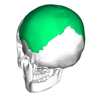
The parietal bones are two bones in the skull which, when joined at a fibrous joint known as a cranial suture, form the sides and roof of the neurocranium. In humans, each bone is roughly quadrilateral in form, and has two surfaces, four borders, and four angles. It is named from the Latin paries (-ietis), wall.

The occipital lobe is one of the four major lobes of the cerebral cortex in the brain of mammals. The name derives from its position at the back of the head, from the Latin ob, 'behind', and caput, 'head'.
Parietal is an adjective used predominantly for the parietal lobe and other relevant anatomy

The lateral ventricles are the two largest ventricles of the brain and contain cerebrospinal fluid. Each cerebral hemisphere contains a lateral ventricle, known as the left or right lateral ventricle, respectively.
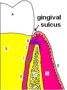
In biological morphology and anatomy, a sulcus is a furrow or fissure. It may be a groove, natural division, deep furrow, elongated cleft, or tear in the surface of a limb or an organ, most notably on the surface of the brain, but also in the lungs, certain muscles, as well as in bones, and elsewhere. Many sulci are the product of a surface fold or junction, such as in the gums, where they fold around the neck of the tooth.

The middle cerebral artery (MCA) is one of the three major paired cerebral arteries that supply blood to the cerebrum. The MCA arises from the internal carotid artery and continues into the lateral sulcus where it then branches and projects to many parts of the lateral cerebral cortex. It also supplies blood to the anterior temporal lobes and the insular cortices.

The lobes of the brain are the four major identifiable regions of the human cerebral cortex, and they comprise the surface of each hemisphere of the cerebrum. The two hemispheres are roughly symmetrical in structure, and are connected by the corpus callosum. Some sources include the insula and limbic lobe but the limbic lobe incorporates parts of the other lobes. The lobes are large areas that are anatomically distinguishable, and are also functionally distinct. Each lobe of the brain has numerous ridges, or gyri, and furrows, sulci that constitute further subzones of the cortex. The expression "lobes of the brain" usually refers only to those of the cerebrum, not to the distinct areas of the cerebellum.
The posterior cerebral artery (PCA) is one of a pair of cerebral arteries that supply oxygenated blood to the occipital lobe, part of the back of the human brain. The two arteries originate from the distal end of the basilar artery, where it bifurcates into the left and right posterior cerebral arteries. These anastomose with the middle cerebral arteries and internal carotid arteries via the posterior communicating arteries.

In human brain anatomy, an operculum, may refer to the frontal, temporal, or parietal operculum, which together cover the insula as the opercula of insula. It can also refer to the occipital operculum, part of the occipital lobe.

The superior parietal lobule is bounded in front by the upper part of the postcentral sulcus, but is usually connected with the postcentral gyrus above the end of the sulcus. The superior parietal lobule contains Brodmann's areas 5 and 7.

The greater wing of the sphenoid bone, or alisphenoid, is a bony process of the sphenoid bone, positioned in the skull behind each eye. There is one on each side, extending from the side of the body of the sphenoid and curving upward, laterally, and backward.

The transverse sinuses, within the human head, are two areas beneath the brain which allow blood to drain from the back of the head. They run laterally in a groove along the interior surface of the occipital bone. They drain from the confluence of sinuses to the sigmoid sinuses, which ultimately connect to the internal jugular vein. See diagram : labeled under the brain as "SIN. TRANS.".

The mastoid part of the temporal bone is the posterior (back) part of the temporal bone, one of the bones of the skull. Its rough surface gives attachment to various muscles and it has openings for blood vessels. From its borders, the mastoid part articulates with two other bones.

The petrous part of the temporal bone is pyramid-shaped and is wedged in at the base of the skull between the sphenoid and occipital bones. Directed medially, forward, and a little upward, it presents a base, an apex, three surfaces, and three angles, and houses in its interior the components of the inner ear. The petrous portion is among the most basal elements of the skull and forms part of the endocranium. Petrous comes from the Latin word petrosus, meaning "stone-like, hard". It is one of the densest bones in the body. In other mammals, it is a separate bone, the petrosal bone.
The tympanic part of the temporal bone is a curved plate of bone lying below the squamous part of the temporal bone, in front of the mastoid process, and surrounding the external part of the ear canal.

The squamous part of occipital bone is situated above and behind the foramen magnum, and is curved from above downward and from side to side.

The body of the sphenoid bone, more or less cubical in shape, is hollowed out in its interior to form two large cavities, the sphenoidal sinuses, which are separated from each other by a septum.

The occipital gyri (OcG) are three gyri in parallel, along the lateral portion of the occipital lobe, also referred to as a composite structure in the brain. The gyri are the superior occipital gyrus, the middle occipital gyrus, and the inferior occipital gyrus, and these are also known as the occipital face area. The superior and inferior occipital sulci separates the three occipital gyri.
