Angular gyrus

Last updated
Angular gyrus
Brain Surface Gyri.SVG
  Angular gyrus
Gray1197.png
Drawing of a cast to illustrate the relations of the brain to the skull. (Angular gyrus labeled at upper left, in yellow section.)
Details
Identifiers
Latin gyrus angularis
NeuroNames 109
NeuroLex ID birnlex_1376
TA98 A14.1.09.124
TA2 5472
FMA 61898
Anatomical terms of neuroanatomy
In this image, the angular gyrus is denoted by the double asterisk ** ParietCapts lateral.png
In this image, the angular gyrus is denoted by the double asterisk **

The angular gyrus is a region of the brain lying mainly in the posteroinferior region of the parietal lobe, occupying the posterior part of the inferior parietal lobule. [1] It represents the Brodmann area 39. [1]

Contents

Its significance is in transferring visual information to Wernicke's area, in order to make meaning out of visually perceived words. [2] It is also involved in a number of processes related to language, number processing and spatial cognition, memory retrieval, attention, and theory of mind.
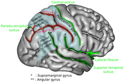
Anatomy

Connections

Left and right angular gyri are connected by the dorsal splenium and isthmus of the corpus callosum. [3]

Connections To the Angular gyrus
Connected To TheVia the
ipsilateral frontal and caudallateral prefrontal and inferior frontal regions superior longitudinal fasciculus. [4]
caudate inferior occipitofrontal fasciculus [5]
parahippocampal gyrus [6] and hippocampus [5] inferior longitudinal fasciculus
precuneus and superior frontal gyrus occipitofrontal fasciculus, [7]
supramarginal gyrus local arcuate [8]

Boundaries

Function

The angular gyrus is the part of the brain associated with complex language functions (i.e. reading, writing and interpretation of what is written). Lesion to this part of the brain shows symptoms of the Gerstmann syndrome: effects include finger agnosia, alexia (inability to read), acalculia (inability to use arithmetic operations), agraphia (inability to copy), and left-right confusion.

Language

Geschwind proposed that written word is translated to internal monologue via the angular gyrus.[ citation needed ]

V. S. Ramachandran, and Edward Hubbard published a paper in 2003 in which they hypothesized the angular gyrus to play a role in understanding metaphors. They stated:

There may be neurological disorders that disturb metaphor and synaesthesia. This has not been studied in detail but we have seen disturbances in the Bouba/Kiki effect (Ramachandran & Hubbard, 2001a) as well as with proverbs in patients with angular gyrus lesions. It would be interesting to see whether they have deficits in other types of synaesthetic metaphor, e.g. 'sharp cheese' or 'loud shirt'. There are also hints that patients with right hemisphere lesions show problems with metaphor. It is possible that their deficits are mainly with spatial metaphors, such as 'He stepped down as director'. [10]

The fact that the angular gyrus is proportionately much larger in hominids than other primates, and its strategic location at the crossroads of areas specialized for processing touch, hearing and vision, leads Ramachandran to believe that it is critical both to conceptual metaphors and to cross-modal abstractions more generally. However, recent research challenges this theory.

Research by Krish Sathian (Emory University) using functional magnetic resonance imaging (fMRI) suggests that the angular gyrus does not play a role in creating conceptual metaphors. Sathian theorizes that conceptual metaphors activate the texture-selective somatosensory cortex in the parietal operculum. [11]

Brownsett and Wise highlight the role of the left angular gyrus in both speaking and writing. [12]

Arithmetic and spatial cognition

Since 1919, brain injuries to the angular gyrus have been known to often cause arithmetic deficits. [13] [14] Functional imaging has shown that while other parts of the parietal lobe bilaterally are involved in approximate calculations due to its link with spatiovisual abilities, the left angular gyrus together with left Inferior frontal gyrus are involved in exact calculation due to verbal arithmetic fact retrieval. [15] When activation in the left angular gyrus is greater, a person's arithmetic skills are also more competent. [16]

Attention

The right angular gyrus has been associated with spatiovisual attention toward salient features. [17] [18] It may allocate attention by employing a bottom-up strategy which draws on the area's ability to attend to retrieved memories. [17] For example, the angular gyrus plays a critical role in distinguishing left from right by integrating the conceptual understanding of the language term "left" or "right" with its location in space. [19] Furthermore, the angular gyrus has been associated with orienting in three dimensional space, not because it interprets space, but because it may control attention shifts in space. [20]

Other functions

Default mode network

The angular gyrus is part of the default mode network, a network of brain regions activated during multi-modal activities that are independent of external stimuli. [21] [22]

Awareness

The angular gyrus reacts differently to intended and consequential movement. [23] This suggests that the angular gyrus monitors the self's intended movements and uses the added information to compute differently, as it does for consequential movements. By recording the discrepancy, the angular gyrus maintains an awareness of the self.

Memory retrieval

Activation of the angular gyrus shows that not only does it mediate memory retrieval, but it also notes contradictions between what is expected from the retrieval, and what is unusual. [3] The angular gyrus can access both content and episodic memories and is useful in inferring from these the intentions of human characters. [17] Furthermore, the angular gyrus may use a feedback strategy to ascertain whether a retrieval is expected or unusual.

Out-of-body experiences

Experiments have demonstrated the ability of stimulation of the right angular gyrus to induce out-of-body experiences. [24] Stimulation of the left angular gyrus in one experiment caused a woman to perceive a shadowy person lurking behind her. The shadowy figure is actually a perceived double of the self. [25] Another such experiment gave the test subject the sensation of being on the ceiling. This is attributed to a discrepancy in the actual position of the body, and the mind's perceived location of the body.

Clinical significance

Damage to the angular gyrus manifests as Gerstmann syndrome. Damage may impair one or more of the below functions.

  1. Dysgraphia/agraphia: deficiency in the ability to write [26] [27]
  2. Dyscalculia/acalculia: difficulty in learning or comprehending mathematics [26] [27]
  3. Finger agnosia: inability to distinguish the fingers on the hand [26] [27]
  4. Left-right disorientation [26] [27]

Additional images

See also

Related Research Articles

Agraphia is an acquired neurological disorder causing a loss in the ability to communicate through writing, either due to some form of motor dysfunction or an inability to spell. The loss of writing ability may present with other language or neurological disorders; disorders appearing commonly with agraphia are alexia, aphasia, dysarthria, agnosia, acalculia and apraxia. The study of individuals with agraphia may provide more information about the pathways involved in writing, both language related and motoric. Agraphia cannot be directly treated, but individuals can learn techniques to help regain and rehabilitate some of their previous writing abilities. These techniques differ depending on the type of agraphia.

<span class="mw-page-title-main">Parietal lobe</span> Part of the brain responsible for sensory input and some language processing

The parietal lobe is one of the four major lobes of the cerebral cortex in the brain of mammals. The parietal lobe is positioned above the temporal lobe and behind the frontal lobe and central sulcus.

<span class="mw-page-title-main">Precuneus</span> Region of the parietal lobe of the brain

In neuroanatomy, the precuneus is the portion of the superior parietal lobule on the medial surface of each brain hemisphere. It is located in front of the cuneus. The precuneus is bounded in front by the marginal branch of the cingulate sulcus, at the rear by the parieto-occipital sulcus, and underneath by the subparietal sulcus. It is involved with episodic memory, visuospatial processing, reflections upon self, and aspects of consciousness.

<span class="mw-page-title-main">Brodmann area 44</span> Brain area

Brodmann area 44, or BA44, is part of the frontal cortex in the human brain. Situated just anterior to premotor cortex (BA6) and on the lateral surface, inferior to BA9.

<span class="mw-page-title-main">Fusiform gyrus</span> Gyrus of the temporal and occipital lobes of the brain

The fusiform gyrus, also known as the lateral occipitotemporal gyrus,is part of the temporal lobe and occipital lobe in Brodmann area 37. The fusiform gyrus is located between the lingual gyrus and parahippocampal gyrus above, and the inferior temporal gyrus below. Though the functionality of the fusiform gyrus is not fully understood, it has been linked with various neural pathways related to recognition. Additionally, it has been linked to various neurological phenomena such as synesthesia, dyslexia, and prosopagnosia.

<span class="mw-page-title-main">Inferior frontal gyrus</span> Part of the brains prefrontal cortex

The inferior frontal gyrus (IFG),, is the lowest positioned gyrus of the frontal gyri, of the frontal lobe, and is part of the prefrontal cortex.

<span class="mw-page-title-main">Transverse temporal gyrus</span> Gyrus of the primary auditory cortex of the brain

The transverse temporal gyri, also called Heschl's gyri or Heschl's convolutions, are gyri found in the area of primary auditory cortex buried within the lateral sulcus of the human brain, occupying Brodmann areas 41 and 42. Transverse temporal gyri are superior to and separated from the planum temporale by Heschl's sulcus. Transverse temporal gyri are found in varying numbers in both the right and left hemispheres of the brain and one study found that this number is not related to the hemisphere or dominance of hemisphere studied in subjects. Transverse temporal gyri can be viewed in the sagittal plane as either an omega shape or a heart shape.

<span class="mw-page-title-main">Insular cortex</span> Portion of the mammalian cerebral cortex

The insular cortex is a portion of the cerebral cortex folded deep within the lateral sulcus within each hemisphere of the mammalian brain.

<span class="mw-page-title-main">Language processing in the brain</span> How humans use words to communicate

In psycholinguistics, language processing refers to the way humans use words to communicate ideas and feelings, and how such communications are processed and understood. Language processing is considered to be a uniquely human ability that is not produced with the same grammatical understanding or systematicity in even human's closest primate relatives.

Acalculia is an acquired impairment in which people have difficulty performing simple mathematical tasks, such as adding, subtracting, multiplying, and even simply stating which of two numbers is larger. Acalculia is distinguished from dyscalculia in that acalculia is acquired late in life due to neurological injury such as stroke, while dyscalculia is a specific developmental disorder first observed during the acquisition of mathematical knowledge. The name comes from the Greek a- meaning "not" and Latin calculare, which means "to count".

<span class="mw-page-title-main">Middle temporal gyrus</span> One of three gyri of the temporal lobe of the brain

Middle temporal gyrus is a gyrus in the brain on the temporal lobe. It is located between the superior temporal gyrus and inferior temporal gyrus. It corresponds largely to Brodmann area 21.

<span class="mw-page-title-main">Superior parietal lobule</span>

The superior parietal lobule is bounded in front by the upper part of the postcentral sulcus, but is usually connected with the postcentral gyrus above the end of the sulcus. The superior parietal lobule contains Brodmann's areas 5 and 7.

<span class="mw-page-title-main">Inferior parietal lobule</span> Portion of the parietal lobe of the brain

The inferior parietal lobule lies below the horizontal portion of the intraparietal sulcus, and behind the lower part of the postcentral sulcus. Also known as Geschwind's territory after Norman Geschwind, an American neurologist, who in the early 1960s recognised its importance. It is a part of the parietal lobe.

<span class="mw-page-title-main">Lingual gyrus</span> Gyrus of the occipital lobe of the brain

The lingual gyrus, also known as the medialoccipitotemporal gyrus, is a brain structure that is linked to processing vision, especially related to letters. It is thought to also play a role in analysis of logical conditions and encoding visual memories. It is named after its shape, which is somewhat similar to a tongue. Contrary to the name, the region has little to do with speech.

<span class="mw-page-title-main">Superior longitudinal fasciculus</span> Association fiber tract of the brain

The superior longitudinal fasciculus (SLF) is an association tract in the brain that is composed of three separate components. It is present in both hemispheres and can be found lateral to the centrum semiovale and connects the frontal, occipital, parietal, and temporal lobes. This bundle of tracts (fasciculus) passes from the frontal lobe through the operculum to the posterior end of the lateral sulcus where they either radiate to and synapse on neurons in the occipital lobe, or turn downward and forward around the putamen and then radiate to and synapse on neurons in anterior portions of the temporal lobe.

<span class="mw-page-title-main">Posterior parietal cortex</span>

The posterior parietal cortex plays an important role in planned movements, spatial reasoning, and attention.

<span class="mw-page-title-main">Superior temporal sulcus</span> Part of the brains temporal lobe

In the human brain, the superior temporal sulcus (STS) is the sulcus separating the superior temporal gyrus from the middle temporal gyrus in the temporal lobe of the brain. A sulcus is a deep groove that curves into the largest part of the brain, the cerebrum, and a gyrus is a ridge that curves outward of the cerebrum.

<span class="mw-page-title-main">Gerstmann syndrome</span> Neuropsychological disorder caused by damage to the inferior parietal lobule

Gerstmann syndrome is a neuropsychological disorder that is characterized by a constellation of symptoms that suggests the presence of a lesion usually near the junction of the temporal and parietal lobes at or near the angular gyrus. Gerstmann syndrome is typically associated with damage to the inferior parietal lobule of the dominant hemisphere. It is classically considered a left-hemisphere disorder, although right-hemisphere damage has also been associated with components of the syndrome.

Finger agnosia, first defined in 1924 by Josef Gerstmann, is the loss in the ability to distinguish, name, or recognize the fingers—not only the patient's own fingers, but also the fingers of others, and drawings and other representations of fingers. It is one of a tetrad of symptoms in Gerstmann syndrome, although it is also possible for finger agnosia to exist on its own without any other disorders. Usually, lesions to the left angular gyrus and posterior parietal areas can lead to finger agnosia.

<span class="mw-page-title-main">Ventrolateral prefrontal cortex</span> Part of the prefrontal cortex of the brain

The ventrolateral prefrontal cortex (VLPFC) is a section of the prefrontal cortex located on the inferior frontal gyrus, bounded superiorly by the inferior frontal sulcus and inferiorly by the lateral sulcus. It is attributed to the anatomical structures of Brodmann's area (BA) 47, 45 and 44.

References

  1. 1 2 Seghier, Mohamed L. (February 2013). "The Angular Gyrus". The Neuroscientist. 19 (1): 43–61. doi:10.1177/1073858412440596. ISSN   1073-8584. PMC   4107834 . PMID   22547530.
  2. John, Hall (2010). Guyton and Hall Textbook of Medical Physiology. Saunders. p. 699. ISBN   978-1416045748.
  3. 1 2 Park, HJ; Kim, JJ; Lee, SK; Seok, JH; Chun, J; Kim, DI; et al. (2008). "Corpus callosal connection mapping using cortical gray matter parcellation and DT-MRI". Human Brain Mapping. 29 (5): 503–16. doi:10.1002/hbm.20314. PMC   6870924 . PMID   17133394.
  4. Makris, Nikos; Kennedy, David N.; McInerney, Sean; Sorensen, A. Gregory; Wang, Ruopeng; Verne, S. Caviness Jr; Pandya, Deepak N. (2005). "Segmentation of Subcomponents within the Superior Longitudinal Fascicle in Humans: A Quantitative, In Vivo, DT-MRI Study". Cereb. Cortex. 15 (6): 854–869. doi: 10.1093/cercor/bhh186 . PMID   15590909.
  5. 1 2 Uddin, Lucina Q.; Supekar, Kaustubh; Amin, Hitha; Rykhlevskaia, Elena; Nguyen, Daniel A.; Greicius, Michael D.; Menon, Vinod (2010). "Dissociable Connectivity within Human Angular Gyrus and Intraparietal Sulcus: Evidence from Functional and Structural Connectivity". Cereb. Cortex. 20 (11): 2636–2646. doi:10.1093/cercor/bhq011. PMC   2951845 . PMID   20154013.
  6. Rushworth, MF; Behrens, TE; Johansen-Berg, H (2006). "Connection patterns distinguish 3 regions of human parietal cortex". Cereb Cortex. 16 (10): 1418–1430. CiteSeerX   10.1.1.504.2825 . doi:10.1093/cercor/bhj079. PMID   16306320.
  7. Makris, Nikos; Papadimitriou, George M.; Sorg, Scott; Kennedy, David N.; Caviness, Verne S.; Pandya, Deepak N. (2007). "The occipitofrontal fascicle in humans: A quantitative, in vivo, DT-MRI study". NeuroImage. 37 (4): 1100–1111. doi:10.1016/j.neuroimage.2007.05.042. PMC   3769215 . PMID   17681797.
  8. Lee, H; Devlin, JT; Shakeshaft, C; Stewart, LH; Brennan, A; Glensman, J; Pitcher, K; Crinion, J; Mechelli, A; Frackowiak, RS; Green, DW; Price, CJ (2007). "Anatomical traces of vocabulary acquisition in the adolescent brain" (PDF). J Neurosci. 27 (5): 1184–1189. doi:10.1523/jneurosci.4442-06.2007. PMC   6673201 . PMID   17267574.
  9. Gai, Dayu. "Angular gyrus | Radiology Reference Article | Radiopaedia.org". Radiopaedia. doi: 10.53347/rid-38811 . Retrieved 2022-06-15.
  10. Ramachandran, V.S.; Hubbard, E.M (2003). "The Phenomenology of Synaesthesia" (PDF). Journal of Consciousness Studies. 10 (8): 49–57.
  11. Simon, K; Stilla, R; Sathian, K (2011). "Metaphorically feeling:Comprehending textual metaphors actives somatosensory cortex". Brain and Language. 120 (3): 416–421. doi:10.1016/j.bandl.2011.12.016. PMC   3318916 . PMID   22305051.
  12. Brownsett, Sonia L. E.; Wise, Richard J. S. (2009-06-16). "The Contribution of the Parietal Lobes to Speaking and Writing". Cerebral Cortex. 20 (3): 517–523. doi:10.1093/cercor/bhp120. ISSN   1460-2199. PMC   2820696 . PMID   19531538.
  13. Henschen, SL (1919). "On language, music and calculation mechanisms and their localisation in the cerebrum". Zeitschrift für die Gesamte Neurologie und Psychiatrie. 52: 273–298. doi:10.1007/bf02872428. S2CID   123138541.
  14. Gerstmann, J (1940). "Syndrome of finger agnosia, disorientation for right and left, agraphia and acalculia—Local diagnostic value". Arch Neurol Psychiatry. 44 (2): 398–408. doi:10.1001/archneurpsyc.1940.02280080158009.
  15. Dehaene, S; Spelke, E; Pinel, P; Stanescu, R; Tsivkin, S (1999). "Sources of mathematical thinking: behavioral and brain-imaging evidence". Science. 284 (5416): 970–4. Bibcode:1999Sci...284..970D. doi:10.1126/science.284.5416.970. PMID   10320379.
  16. Grabner, RH; Ansari, D; Reishofer, G; Stern, E; Ebner, F; Neuper, C (2007). "Individual differences in mathematical competence predict parietal brain activation during mental calculation". NeuroImage. 38 (2): 346–56. doi:10.1016/j.neuroimage.2007.07.041. PMID   17851092. S2CID   18960661.
  17. 1 2 3 Seghier, M. L. (2012). "The angular gyrus: multiple function ad multiple subdivisions". Neuroscientist. 19 (1): 43–61. doi:10.1177/1073858412440596. PMC   4107834 . PMID   22547530.
  18. Arsalidou, M; Taylor, MJ (2011). "Is 2+2=4? Meta-analyses of brain areas needed for numbers and calculations". NeuroImage. 54 (3): 2382–93. doi:10.1016/j.neuroimage.2010.10.009. PMID   20946958. S2CID   140205603.
  19. Hirnstein, M; Bayer, U; Ellison, A; Hausmann, M (2011). "TMS over the left angular gyrus impairs the ability to discriminate left from right". Neuropsychologia. 49 (1): 29–33. doi:10.1016/j.neuropsychologia.2010.10.028. PMID   21035475. S2CID   8460451.
  20. Chen, Q; Weidner, R; Vossel, S; Weiss, PH; Fink, GR (Sep 2012). "Neural mechanisms of attentional reorienting in three-dimensional space". J Neurosci. 32 (39): 13352–62. doi: 10.1523/jneurosci.1772-12.2012 . PMC   6621370 . PMID   23015426.
  21. Murphy, Charlotte; Jefferies, Elizabeth; Rueschemeyer, Shirley-Ann; Sormaz, Mladen; Wang, Hao-ting; Margulies, Daniel S.; Smallwood, Jonathan (2018-05-01). "Distant from input: Evidence of regions within the default mode network supporting perceptually-decoupled and conceptually-guided cognition". NeuroImage. 171: 393–401. doi: 10.1016/j.neuroimage.2018.01.017 . ISSN   1053-8119. PMC   5883322 . PMID   29339310.
  22. Vatansever, D.; Manktelow, A. E.; Sahakian, B. J.; Menon, D. K.; Stamatakis, E. A. (January 2017). "Angular default mode network connectivity across working memory load". Human Brain Mapping. 38 (1): 41–52. doi:10.1002/hbm.23341. ISSN   1097-0193. PMC   6866899 . PMID   27489137.
  23. Farrer C, Frey SH, Van Horn JD, Tunik E, Turk D, Inati S, Grafton ST. The angular gyrus computes action awareness representations. Centre de Neuroscience Cognitive.
  24. Out-of-Body Experience? Your Brain Is to Blame - New York Times
  25. Arzy, S.; Seeck, M.; Ortigue, S.; Spinelli, L.; Blanke, O. (2006). "Induction of an illusory shadow person: Stimulation of a site on the brain's left hemisphere prompts the creepy feeling that somebody is close by". Nature. 443 (21): 287. doi: 10.1038/443287a . PMID   16988702. S2CID   4338465.
  26. 1 2 3 4 Vallar G (July 2007). "Spatial neglect, Balint-Homes' and Gerstmann's syndrome, and other spatial disorders". CNS Spectr. 12 (7): 527–36. doi:10.1017/S1092852900021271. PMID   17603404. S2CID   45201083.
  27. 1 2 3 4 Carota A, Di Pietro M, Ptak R, Poglia D, Schnider A (2004). "Defective spatial imagery with pure Gerstmann's syndrome". Eur. Neurol. 52 (1): 1–6. doi:10.1159/000079251. PMID   15218337. S2CID   22897847.