Gerstmann syndrome

Last updated
Gerstmann syndrome
Gray726 inferior parietal lobule.png
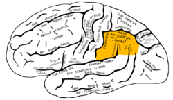
The inferior parietal lobule, with damage in this area most often associated with Gerstmann syndrome
Specialty Neurology, neuropsychology   OOjs UI icon edit-ltr-progressive.svg
Symptoms Dysgraphia, dyscalculia, finger agnosia, left-right disorientation, constructional apraxia, aphasia
Causes Idiopathic, stroke, dementia

Gerstmann syndrome is a neuropsychological disorder that is characterized by a constellation of symptoms [1] that suggests the presence of a lesion usually near the junction of the temporal and parietal lobes at or near the angular gyrus. Gerstmann syndrome is typically associated with damage to the inferior parietal lobule of the dominant hemisphere. It is classically considered a left-hemisphere disorder, although right-hemisphere damage has also been associated with components of the syndrome. [2]

Contents

It is named after Jewish Austrian-born American neurologist Josef Gerstmann. [3]

Symptoms

Gerstmann syndrome is characterized by four primary symptoms, collectively referred to as a tetrad:

  1. Dysgraphia/agraphia: deficiency in the ability to write [4] [5]
  2. Dyscalculia/acalculia: difficulty in learning or comprehending mathematics [4] [5]
  3. Finger agnosia: inability to distinguish the fingers on the hand [4] [5]
  4. Left-right disorientation [4] [5]

Causes

This disorder is often associated with brain lesions in the dominant (usually left) hemisphere including the angular and supramarginal gyri (Brodmann area 39 and 40 respectively) near the temporal and parietal lobe junction. There is significant debate in the scientific literature as to whether Gerstmann syndrome truly represents a unified, theoretically motivated syndrome. Thus its diagnostic utility has been questioned by neurologists and neuropsychologists alike. The angular gyrus is generally involved in translating visual patterns of letters and words into meaningful information, such as is done while reading.[ citation needed ]

In adults

In adults, the syndrome may occur after a stroke. [5] In addition to exhibiting the above symptoms, many adults also experience dysphasia or aphasia, [2] which is difficulty in expressing oneself when speaking, in understanding speech, or in reading and writing.[ citation needed ]

In children

There are few reports of the syndrome, sometimes called developmental Gerstmann syndrome, in children. [6] The cause is not known. Most cases are identified when children reach school age, a time when they are challenged with writing and mathematic exercises. Generally, children with the disorder exhibit poor handwriting and spelling skills, and difficulty with math functions, including adding, subtracting, multiplying, and dividing. An inability to differentiate right from left and to discriminate among individual fingers may also be apparent. In addition to the four primary symptoms, many children also have constructional apraxia, an inability to copy simple drawings. Frequently, there is also an impairment in reading. Children at any level of intelligence may be affected with the disorder. [6]

Diagnosis

Diagnosis may be clinical if associated with dementia and other etiologies. In cases caused by stroke, MRI will show a corresponding stroke in the inferior parietal lobule. In the acute stage, this will be bright (restricted diffusion) on the DWI sequence and dark at the corresponding area on the ADC sequence.[ vague ][ citation needed ]

Treatment

There is no cure for Gerstmann syndrome. Treatment is symptomatic and supportive. Occupational and speech therapies may help diminish the dysgraphia and apraxia. In addition, calculators and word processors may help school children cope with the symptoms of the disorder. [7]

Prognosis

In adults, many of the symptoms diminish over time. Although it has been suggested that a similar diminishing of symptoms occurs in children as well, it appears more likely that most do not overcome their deficits, but instead simply learn to adjust. [7]

See also

Related Research Articles

<span class="mw-page-title-main">Apraxia</span> Medical condition

Apraxia is a motor disorder caused by damage to the brain, which causes difficulty with motor planning to perform tasks or movements. The nature of the damage determines the disorder's severity, and the absence of sensory loss or paralysis helps to explain the level of difficulty. Children may be born with apraxia; its cause is unknown, and symptoms are usually noticed in the early stages of development. Apraxia occurring later in life, known as acquired apraxia, is typically caused by traumatic brain injury, stroke, dementia, Alzheimer's disease, brain tumor, or other neurodegenerative disorders. The multiple types of apraxia are categorized by the specific ability and/or body part affected.

Agraphia is an acquired neurological disorder causing a loss in the ability to communicate through writing, either due to some form of motor dysfunction or an inability to spell. The loss of writing ability may present with other language or neurological disorders; disorders appearing commonly with agraphia are alexia, aphasia, dysarthria, agnosia, acalculia and apraxia. The study of individuals with agraphia may provide more information about the pathways involved in writing, both language related and motoric. Agraphia cannot be directly treated, but individuals can learn techniques to help regain and rehabilitate some of their previous writing abilities. These techniques differ depending on the type of agraphia.

<span class="mw-page-title-main">Brain damage</span> Destruction or degeneration of brain cells

Neurotrauma, brain damage or brain injury (BI) is the destruction or degeneration of brain cells. Brain injuries occur due to a wide range of internal and external factors. In general, brain damage refers to significant, undiscriminating trauma-induced damage.

<span class="mw-page-title-main">Parietal lobe</span> Part of the brain responsible for sensory input and some language processing

The parietal lobe is one of the four major lobes of the cerebral cortex in the brain of mammals. The parietal lobe is positioned above the temporal lobe and behind the frontal lobe and central sulcus.

Dyscalculia is a disability resulting in difficulty learning or comprehending arithmetic, such as difficulty in understanding numbers, learning how to manipulate numbers, performing mathematical calculations, and learning facts in mathematics. It is sometimes colloquially referred to as "math dyslexia", though this analogy is misleading as they are distinct syndromes.

Dysgraphia is a neurological disorder and learning disability that concerns written expression, which affects the ability to write, primarily handwriting, but also coherence. It is a specific learning disability (SLD) as well as a transcription disability, meaning that it is a writing disorder associated with impaired handwriting, orthographic coding and finger sequencing. It often overlaps with other learning disabilities and neurodevelopmental disorders such as speech impairment, attention deficit hyperactivity disorder (ADHD) or developmental coordination disorder (DCD).

<span class="mw-page-title-main">Bálint's syndrome</span> Medical condition

Bálint's syndrome is an uncommon and incompletely understood triad of severe neuropsychological impairments: inability to perceive the visual field as a whole (simultanagnosia), difficulty in fixating the eyes, and inability to move the hand to a specific object by using vision. It was named in 1909 for the Austro-Hungarian neurologist and psychiatrist Rezső Bálint who first identified it.

<span class="mw-page-title-main">Angular gyrus</span> Gyrus of the parietal lobe of the brain

The angular gyrus is a region of the brain lying mainly in the posteroinferior region of the parietal lobe, occupying the posterior part of the inferior parietal lobule. It represents the Brodmann area 39.

Acalculia is an acquired impairment in which people have difficulty performing simple mathematical tasks, such as adding, subtracting, multiplying, and even simply stating which of two numbers is larger. Acalculia is distinguished from dyscalculia in that acalculia is acquired late in life due to neurological injury such as stroke, while dyscalculia is a specific developmental disorder first observed during the acquisition of mathematical knowledge. The name comes from the Greek a- meaning "not" and Latin calculare, which means "to count".

<span class="mw-page-title-main">Frontal lobe disorder</span> Brain disorder

Frontal lobe disorder, also frontal lobe syndrome, is an impairment of the frontal lobe of the brain due to disease or frontal lobe injury. The frontal lobe plays a key role in executive functions such as motivation, planning, social behaviour, and speech production. Frontal lobe syndrome can be caused by a range of conditions including head trauma, tumours, neurodegenerative diseases, neurodevelopmental disorders, neurosurgery and cerebrovascular disease. Frontal lobe impairment can be detected by recognition of typical signs and symptoms, use of simple screening tests, and specialist neurological testing.

<span class="mw-page-title-main">Inferior parietal lobule</span> Portion of the parietal lobe of the brain

The inferior parietal lobule lies below the horizontal portion of the intraparietal sulcus, and behind the lower part of the postcentral sulcus. Also known as Geschwind's territory after Norman Geschwind, an American neurologist, who in the early 1960s recognised its importance. It is a part of the parietal lobe.

<span class="mw-page-title-main">Foix–Chavany–Marie syndrome</span> Medical condition

Foix–Chavany–Marie Syndrome (FCMS), also known as bilateral opercular syndrome, is a neuropathological disorder characterized by paralysis of the facial, tongue, pharynx, and masticatory muscles of the mouth that aid in chewing. The disorder is primarily caused by thrombotic and embolic strokes, which cause a deficiency of oxygen in the brain. As a result, bilateral lesions may form in the junctions between the frontal lobe and temporal lobe, the parietal lobe and cortical lobe, or the subcortical region of the brain. FCMS may also arise from defects existing at birth that may be inherited or nonhereditary. Symptoms of FCMS can be present in a person of any age and it is diagnosed using automatic-voluntary dissociation assessment, psycholinguistic testing, neuropsychological testing, and brain scanning. Treatment for FCMS depends on the onset, as well as on the severity of symptoms, and it involves a multidisciplinary approach.

Focal neurologic signs also known as focal neurological deficits or focal CNS signs are impairments of nerve, spinal cord, or brain function that affects a specific region of the body, e.g. weakness in the left arm, the right leg, paresis, or plegia.

Autotopagnosia from the Greek a and gnosis, meaning "without knowledge", topos meaning "place", and auto meaning "oneself", autotopagnosia virtually translates to the "lack of knowledge about one's own space," and is clinically described as such.

Ideomotor Apraxia, often IMA, is a neurological disorder characterized by the inability to correctly imitate hand gestures and voluntarily mime tool use, e.g. pretend to brush one's hair. The ability to spontaneously use tools, such as brushing one's hair in the morning without being instructed to do so, may remain intact, but is often lost. The general concept of apraxia and the classification of ideomotor apraxia were developed in Germany in the late 19th and early 20th centuries by the work of Hugo Liepmann, Adolph Kussmaul, Arnold Pick, Paul Flechsig, Hermann Munk, Carl Nothnagel, Theodor Meynert, and linguist Heymann Steinthal, among others. Ideomotor apraxia was classified as "ideo-kinetic apraxia" by Liepmann due to the apparent dissociation of the idea of the action with its execution. The classifications of the various subtypes are not well defined at present, however, owing to issues of diagnosis and pathophysiology. Ideomotor apraxia is hypothesized to result from a disruption of the system that relates stored tool use and gesture information with the state of the body to produce the proper motor output. This system is thought to be related to the areas of the brain most often seen to be damaged when ideomotor apraxia is present: the left parietal lobe and the premotor cortex. Little can be done at present to reverse the motor deficit seen in ideomotor apraxia, although the extent of dysfunction it induces is not entirely clear.

Amorphosynthesis, also called a hemi-sensory deficit, is a neuropsychological condition in which a patient experiences unilateral inattention to sensory input. This phenomenon is frequently associated with damage to the right cerebral hemisphere resulting in severe sensory deficits that are observed on the contralesional (left) side of the body. A right-sided deficit is less commonly observed and the effects are reported to be temporary and minor. Evidence suggests that the right cerebral hemisphere has a dominant role in attention and awareness to somatic sensations through ipsilateral and contralateral stimulation. In contrast, the left cerebral hemisphere is activated only by contralateral stimuli. Thus, the left and right cerebral hemispheres exhibit redundant processing to the right-side of the body and a lesion to the left cerebral hemisphere can be compensated by the ipsiversive processes of the right cerebral hemisphere. For this reason, right-sided amorphosynthesis is less often observed and is generally associated with bilateral lesions.

Constructional apraxia Apraxia is a neurological disorder in which people are unable to perform tasks or movements even though they understand the task, are willing to complete it, and have the physical ability to perform the movements. It is characterized by an inability or difficulty to build, assemble, or draw objects. Constructional apraxia may be caused by lesions in the parietal lobe following stroke or it may serve as an indicator for Alzheimer's disease.

Visuospatial dysgnosia is a loss of the sense of "whereness" in the relation of oneself to one's environment and in the relation of objects to each other. Visuospatial dysgnosia is often linked with topographical disorientation.

Finger agnosia, first defined in 1924 by Josef Gerstmann, is the loss in the ability to distinguish, name, or recognize the fingers—not only the patient's own fingers, but also the fingers of others, and drawings and other representations of fingers. It is one of a tetrad of symptoms in Gerstmann syndrome, although it is also possible for finger agnosia to exist on its own without any other disorders. Usually, lesions to the left angular gyrus and posterior parietal areas can lead to finger agnosia.

<span class="mw-page-title-main">Disconnection syndrome</span> Collection of neurological symptoms

Disconnection syndrome is a general term for a collection of neurological symptoms caused – via lesions to associational or commissural nerve fibres – by damage to the white matter axons of communication pathways in the cerebrum, independent of any lesions to the cortex. The behavioral effects of such disconnections are relatively predictable in adults. Disconnection syndromes usually reflect circumstances where regions A and B still have their functional specializations except in domains that depend on the interconnections between the two regions.

References

  1. João RB, Filgueiras RM, Mussi ML, de Barros JE (June 2017). "Transient Gerstmann syndrome as manifestation of stroke: Case report and brief literature review". Dementia & Neuropsychologia. 11 (2): 202–205. doi:10.1590/1980-57642016dn11-020013. PMC   5710689 . PMID   29213512.
  2. 1 2 Heimburger RF, Demyer W, Reitan RM (February 1964). "Implications of Gerstmann's syndrome". Journal of Neurology, Neurosurgery, and Psychiatry. 27 (1): 52–57. doi:10.1136/jnnp.27.1.52. PMC   495679 . PMID   14123925.
  3. synd/2267 at Who Named It?
  4. 1 2 3 4 Vallar G (July 2007). "Spatial neglect, Balint-Homes' and Gerstmann's syndrome, and other spatial disorders". CNS Spectrums. 12 (7): 527–536. doi:10.1017/S1092852900021271. PMID   17603404. S2CID   45201083.
  5. 1 2 3 4 5 Carota A, Di Pietro M, Ptak R, Poglia D, Schnider A (2004). "Defective spatial imagery with pure Gerstmann's syndrome". European Neurology. 52 (1): 1–6. doi:10.1159/000079251. PMID   15218337. S2CID   22897847.
  6. 1 2 Miller CJ, Hynd GW (April 2004). "What ever happened to developmental Gerstmann's syndrome? Links to other pediatric, genetic, and neurodevelopmental syndromes". Journal of Child Neurology. 19 (4): 282–289. doi:10.1177/088307380401900408. PMID   15163095. S2CID   26407185.
  7. 1 2 Office of Communications and Public Liaison. "Gerstmann's Syndrome Information". U.S. National Institute of Neurological Disorders and Stroke (NINDS). Bethesda, MD. Archived from the original on 2012-06-24. Retrieved 2012-06-27.

Further reading