Medial pontine syndrome

Last updated
Medial pontine syndrome
Pons section at facial colliculus.png
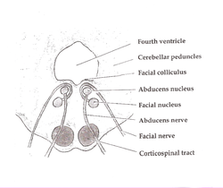
Pons. (Medial pontine syndrome affects structures at the bottom of the diagram: the corticospinal tract, abducens nerve, and occasionally the facial nerve. Medial lemniscus is also affected, but not pictured.)
Specialty Neurology   OOjs UI icon edit-ltr-progressive.svg

Medial inferior pontine syndrome is a condition associated with a contralateral hemiplegia.[ citation needed ]"Medial inferior pontine syndrome" has been described as equivalent to Foville's syndrome. [1]

Contents

Presentation

Although medial pontine syndrome has many similarities to medial medullary syndrome, because it is located higher up the brainstem in the pons, it affects a different set of cranial nuclei.[ citation needed ]

Structure affectedPresentation
Corticospinal tract Contralateral spastic hemiparesis
Medial lemniscus Contralateral PCML (aka DCML) pathway loss (tactile, vibration, and stereognosis)
Abducens nerve Strabismus (ipsilateral lateral rectus muscle paralysis - the affected eye looks down and towards the nose). Abducens nerve lesion localizes the lesion to inferior pons.

Depending upon the size of the infarct, it can also involve the facial nerve.

Cause

Human brainstem blood supply description. Basilar artery is #7, and pons is visible below it. Human brainstem blood supply description.JPG
Human brainstem blood supply description. Basilar artery is #7, and pons is visible below it.

Medial pontine syndrome results from occlusion of paramedian branches of the basilar artery.[ citation needed ]


Treatment

See also

Related Research Articles

<span class="mw-page-title-main">Medulla oblongata</span> Structure of the brain stem

The medulla oblongata or simply medulla is a long stem-like structure which makes up the lower part of the brainstem. It is anterior and partially inferior to the cerebellum. It is a cone-shaped neuronal mass responsible for autonomic (involuntary) functions, ranging from vomiting to sneezing. The medulla contains the cardiovascular center, the respiratory center, vomiting and vasomotor centers, responsible for the autonomic functions of breathing, heart rate and blood pressure as well as the sleep–wake cycle. "Medulla" is from Latin, ‘pith or marrow’. And "oblongata" is from Latin, ‘lengthened or longish or elongated'.

<span class="mw-page-title-main">Abducens nerve</span> Cranial nerve VI, for eye movements

The abducens nerve or abducent nerve, also known as the sixth cranial nerve, cranial nerve VI, or simply CN VI, is a cranial nerve in humans and various other animals that controls the movement of the lateral rectus muscle, one of the extraocular muscles responsible for outward gaze. It is a somatic efferent nerve.

<span class="mw-page-title-main">Brainstem</span> Posterior part of the brain, adjoining and structurally continuous

The brainstem is the posterior stalk-like part of the brain that connects the cerebrum with the spinal cord. In the human brain the brainstem is composed of the midbrain, the pons, and the medulla oblongata. The midbrain is continuous with the thalamus of the diencephalon through the tentorial notch, and sometimes the diencephalon is included in the brainstem.

<span class="mw-page-title-main">Internal capsule</span> White matter structure situated in the inferomedial part of each cerebral hemisphere of the brain

The internal capsule is a paired white matter structure, as a two-way tract, carrying ascending and descending fibers, to and from the cerebral cortex. The internal capsule is situated in the inferomedial part of each cerebral hemisphere of the brain. It carries information past the subcortical basal ganglia. As it courses it separates the caudate nucleus and the thalamus from the putamen and the globus pallidus. It also separates the caudate nucleus and the putamen in the dorsal striatum, a brain region involved in motor and reward pathways.

<span class="mw-page-title-main">Lateral medullary syndrome</span> Medical condition

Lateral medullary syndrome is a neurological disorder causing a range of symptoms due to ischemia in the lateral part of the medulla oblongata in the brainstem. The ischemia is a result of a blockage most commonly in the vertebral artery or the posterior inferior cerebellar artery. Lateral medullary syndrome is also called Wallenberg's syndrome, posterior inferior cerebellar artery (PICA) syndrome and vertebral artery syndrome.

<span class="mw-page-title-main">Medial medullary syndrome</span> Medical condition

Medial medullary syndrome, also known as inferior alternating syndrome, hypoglossal alternating hemiplegia, lower alternating hemiplegia, or Dejerine syndrome, is a type of alternating hemiplegia characterized by a set of clinical features resulting from occlusion of the anterior spinal artery. This results in the infarction of medial part of the medulla oblongata.

<span class="mw-page-title-main">Anterior spinal artery</span> Blood vessel in the front of the spinal cord

In human anatomy, the anterior spinal artery is the artery that supplies the anterior portion of the spinal cord. It arises from branches of the vertebral arteries and courses along the anterior aspect of the spinal cord. It is reinforced by several contributory arteries, especially the artery of Adamkiewicz.

<span class="mw-page-title-main">Ophthalmoparesis</span> Weakness or paralysis of extraocular muscles

Ophthalmoparesis refers to weakness (-paresis) or paralysis (-plegia) of one or more extraocular muscles which are responsible for eye movements. It is a physical finding in certain neurologic, ophthalmologic, and endocrine disease.

Abnormal posturing is an involuntary flexion or extension of the arms and legs, indicating severe brain injury. It occurs when one set of muscles becomes incapacitated while the opposing set is not, and an external stimulus such as pain causes the working set of muscles to contract. The posturing may also occur without a stimulus. Since posturing is an important indicator of the amount of damage that has occurred to the brain, it is used by medical professionals to measure the severity of a coma with the Glasgow Coma Scale and the Pediatric Glasgow Coma Scale.

<span class="mw-page-title-main">Posterior cerebral artery</span> Artery which supplies blood to the occipital lobe of the brain

The posterior cerebral artery (PCA) is one of a pair of cerebral arteries that supply oxygenated blood to the occipital lobe, part of the back of the human brain. The two arteries originate from the distal end of the basilar artery, where it bifurcates into the left and right posterior cerebral arteries. These anastomose with the middle cerebral arteries and internal carotid arteries via the posterior communicating arteries.

<span class="mw-page-title-main">Posterior inferior cerebellar artery</span>

The posterior inferior cerebellar artery (PICA) is the largest branch of the vertebral artery. It is one of the three main arteries that supply blood to the cerebellum, a part of the brain. Blockage of the posterior inferior cerebellar artery can result in a type of stroke called lateral medullary syndrome.

<span class="mw-page-title-main">Sixth nerve palsy</span> Inability to turn out the eye due to dysfunction of the abducens nerve

Sixth nerve palsy, or abducens nerve palsy, is a disorder associated with dysfunction of cranial nerve VI, which is responsible for causing contraction of the lateral rectus muscle to abduct the eye. The inability of an eye to turn outward, results in a convergent strabismus or esotropia of which the primary symptom is diplopia in which the two images appear side-by-side. Thus, the diplopia is horizontal and worse in the distance. Diplopia is also increased on looking to the affected side and is partly caused by overaction of the medial rectus on the unaffected side as it tries to provide the extra innervation to the affected lateral rectus. These two muscles are synergists or "yoke muscles" as both attempt to move the eye over to the left or right. The condition is commonly unilateral but can also occur bilaterally.

<span class="mw-page-title-main">Weber's syndrome</span> Medical condition

Weber's syndrome, also known as midbrain stroke syndrome or superior alternating hemiplegia, is a form of stroke that affects the medial portion of the midbrain. It involves oculomotor fascicles in the interpeduncular cisterns and cerebral peduncle so it characterizes the presence of an ipsilateral lower motor neuron type oculomotor nerve palsy and contralateral hemiparesis or hemiplegia.

<span class="mw-page-title-main">Foville's syndrome</span> Medical condition

Foville's syndrome is caused by the blockage of the perforating branches of the basilar artery in the region of the brainstem known as the pons. It is most frequently caused by lesions such as vascular disease and tumors involving the dorsal pons.

<span class="mw-page-title-main">Medial vestibular nucleus</span>

The medial vestibular nucleus is one of the vestibular nuclei. It is located in the medulla oblongata.

<span class="mw-page-title-main">Lateral pontine syndrome</span> Medical condition

Lateral pontine syndrome, also known as Marie-Foix syndrome or Marie-Foix-Alajouanine syndrome is one of the brainstem stroke syndromes of the lateral aspect of the pons. A lateral pontine syndrome is a lesion which is similar to the lateral medullary syndrome, but because it occurs in the pons, it also involves the cranial nerve nuclei of the pons.

<span class="mw-page-title-main">Juxtarestiform body</span> Subdivision of the Inferior Cerebellar Penduncle

The juxtarestiform body is the smaller, medial subdivision of each inferior cerebellar peduncle.

A brainstem stroke syndrome falls under the broader category of stroke syndromes, or specific symptoms caused by vascular injury to an area of brain. As the brainstem contains numerous cranial nuclei and white matter tracts, a stroke in this area can have a number of unique symptoms depending on the particular blood vessel that was injured and the group of cranial nerves and tracts that are no longer perfused. Symptoms of a brainstem stroke frequently include sudden vertigo and ataxia, with or without weakness. Brainstem stroke can also cause diplopia, slurred speech and decreased level of consciousness. A more serious outcome is locked-in syndrome.

Alternating hemiplegia is a form of hemiplegia that has an ipsilateral cranial nerve palsies and contralateral hemiplegia or hemiparesis of extremities of the body. The disorder is characterized by recurrent episodes of paralysis on one side of the body. There are multiple forms of alternating hemiplegia, Weber's syndrome, middle alternating hemiplegia, and inferior alternating hemiplegia. This type of syndrome can result from a unilateral lesion in the brainstem affecting both upper motor neurons and lower motor neurons. The muscles that would receive signals from these damaged upper motor neurons result in spastic paralysis. With a lesion in the brainstem, this affects the majority of limb and trunk muscles on the contralateral side due to the upper motor neurons decussation after the brainstem. The cranial nerves and cranial nerve nuclei are also located in the brainstem making them susceptible to damage from a brainstem lesion. Cranial nerves III (Oculomotor), VI (Abducens), and XII (Hypoglossal) are most often associated with this syndrome given their close proximity with the pyramidal tract, the location which upper motor neurons are in on their way to the spinal cord. Damages to these structures produce the ipsilateral presentation of paralysis or palsy due to the lack of cranial nerve decussation before innervating their target muscles. The paralysis may be brief or it may last for several days, many times the episodes will resolve after sleep. Some common symptoms of alternating hemiplegia are mental impairment, gait and balance difficulties, excessive sweating and changes in body temperature.

References

  1. Hubloue I, Laureys S, Michotte A (September 1996). "A rare case of diplopia: medial inferior pontine syndrome or Foville's syndrome". Eur J Emerg Med. 3 (3): 194–8. doi:10.1097/00063110-199609000-00011. PMID   9023501.