Posterior cerebral artery | |
---|---|
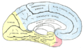
![]() The outer surface of the human brain, with the area supplied by the posterior cerebral artery shown in yellow. | |
![]() The arterial circle and arteries of the brain (inferior view). The posterior cerebral arteries (bottom forks) arise from the basilar artery (center). | |
Details | |
Source | Basilar artery (most common in adults) |
Vein | Cerebral veins |
Supplies | Occipital lobe as well as medial and inferior temporal lobe of cerebrum |
Identifiers | |
Latin | arteria cerebri posterior |
Acronym(s) | PCA |
MeSH | D020769 |
TA98 | A12.2.07.082 |
TA2 | 4565 |
FMA | 50583 |
Anatomical terminology |
The posterior cerebral artery (PCA) is one of a pair of cerebral arteries that supply oxygenated blood to the occipital lobe, as well as the medial and inferior aspects of the temporal lobe of the human brain. The two arteries originate from the distal end of the basilar artery, where it bifurcates into the left and right posterior cerebral arteries. These anastomose with the middle cerebral arteries and internal carotid arteries via the posterior communicating arteries.
The posterior cerebral artery is subdivided into 4 segments:
P1: pre-communicating segment
P2: post-communicating segment
P3: quadrigeminal segment
P4: cortical segment
The branches of the posterior cerebral artery are divided into two sets, ganglionic and cortical.
The following are central branches of the PCA, also known as perforating branches:
The posterior choroidal branches of the posterior cerebral artery are sometimes referred to as a single posterior choroidal artery.
The cortical branches are:
The prenatal development of the posterior cerebral arteries in the fetus comes relatively late and arises from the fusion of several embryonic vessels near the caudal ends of the posterior communicating arteries supplying the mesencephalon and diencephalon. [2] The PCA thus begins as a continuation of the PCommA in the fetus with only 10–30% of fetuses showing a prominent basilar origin. [3]
The fetal carotid origin of the PCA usually regresses as the vertebral and basilar arteries develop with the posterior communicating artery reducing in size. In most adults, the PCA sources from the anterior portion of the basilar artery. About 19% of adults retain PCommA dominance of the PCA, with 72% having dominant basilar origin, and the rest having either equal prominence between PCommA and basilar artery, or a single exclusive source. [3]
Signs and symptoms: Structures involved