Anterior choroidal artery

Last updated

Anterior choroidal artery
Circle of Willis en.svg
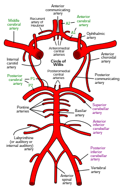
Diagram of the arterial circulation at the base of the brain (inferior view). The anterior choroidal artery is labeled at right of the circle of Willis.
Details
Source Internal carotid artery
Identifiers
Latin arteria choroidea anterior
TA98 A12.2.06.019
TA2 4501
FMA 50087
Anatomical terminology

The anterior choroidal artery is a bilaterally paired artery of the brain. It is typically a branch of the internal carotid artery which supplies the choroid plexus of lateral ventricle and third ventricle as well as numerous structures of the brain.

Contents

Occlusion of the artery can result in loss of sensation, loss of part of the visual field, and impaired movement, all on the opposite side of the body as the occlusion.

Structure

Origin

The anterior choroidal artery typically originates from the internal carotid artery. It may (rarely) instead arise from the middle cerebral artery. [1]

It originates from the distal internal carotid artery (ICA) 5 mm distal to the origin of the posterior communicating artery and just proximal to the terminal bifurcation of the ICA. [2]

Course

It initially course posterolaterally on the inferior surface of the cerebral hemisphere alongside the optic tract, crossing the tract medial-to-lateral [3] inferior to the tract. [4] At the level of the lateral geniculate nucleus, it curves around the lateral aspect of the cerebral peduncle to reach its posterior aspect. It reaches the medial portion of the transverse cerebral fissure to enter the lateral ventricle. [3] It enters the lateral ventricle at the apex of its inferior horn, at the inferior extremity of choroid fissure, just superior to the uncus. [4]

Distribution

It serves structures in the prosencephalon, diencephalon, and mesencephalon: [2]

Clinical significance

Occlusion of the artery results in contralateral hemianopsia (partial loss of vision) and hemianaesthesia (loss of sensation), as well as partial hemiplegia (loss of the ability to move). [4] These symptoms are thought to arise from ischemic damage to the posterior limb of the internal capsule, thalamus, and optic chiasm/optic tract. However, the posterior limb of the internal capsule also receives lenticulostriate arteries from the middle cerebral artery, thus creating partially redundant supply. [5]

See also

References

  1. Thomas Lathrop Stedman (2006). Stedman's medical dictionary. [electronic resource]. Internet Archive. Lippincott Williams & Wilkins. ISBN   978-0-683-40007-6.
  2. 1 2 Ropper, Maurice Victor, Allan H. (2001). Adams and Victor's principles of neurology (7th ed.). New York: Medical Pub. Division, McGraw-Hill. ISBN   0-07-067497-3.{{cite book}}: CS1 maint: multiple names: authors list (link)
  3. 1 2 3 4 "anterior choroidal artery - Dictionnaire médical de l'Académie de Médecine". www.academie-medecine.fr. Retrieved 12 August 2024.
  4. 1 2 3 Sinnatamby, Chummy S. (2011). Last's Anatomy (12th ed.). p. 473. ISBN   978-0-7295-3752-0.
  5. Helgason, C; Caplan, LR (1986). "Anterior choroidal artery-territory infarction: Report of cases and review". Arch. Neurol. 43 (7): 681–686. doi:10.1001/archneur.1986.00520070039015. PMID   3729746.