Transverse cervical artery

Last updated

Transverse cervical artery
Thyrocervical trunk.png
Superficial dissection of the right side of the neck, showing the carotid and subclavian arteries (transverse cervical artery is labeled, branching from the thyrocervical trunk)
Superficial and deep branches.png
Superficial and deep branches from the transverse cervical artery
Details
Source Thyrocervical trunk
Branches Superficial branch
Dorsal scapular artery (Deep Branch)
Vein Transverse cervical veins
SuppliesThe trapezius and Sternocleidomastoid muscles
Identifiers
Latin arteria transversa cervicis,
arteria transversa colli
TA98 A12.2.08.053
TA2 4601
FMA 10664
Anatomical terminology

The transverse cervical artery (transverse artery of neck or transversa colli artery) is an artery in the neck and a branch of the thyrocervical trunk, running at a higher level than the suprascapular artery.

Contents

Structure

It passes transversely below the inferior belly of the omohyoid muscle to the anterior margin of the trapezius, beneath which it divides into a superficial and a deep branch.

It crosses in front of the phrenic nerve and the scalene muscles, and in front of or between the divisions of the brachial plexus, and is covered by the platysma and sternocleidomastoid muscles, and crossed by the omohyoid and trapezius.

The transverse cervical artery originates from the thyrocervical trunk, it passes through the posterior triangle of the neck to the anterior border of the levator scapulae muscle, where it divides into deep and superficial branches.

Function

Superficial branch

Upon entering the trapezius muscle the superficial branch divides again into an ascending and descending branch. The ascending branch distributes branches to trapezius, and to the neighboring muscles and lymph glands in the neck, and anastomoses with the superficial branch of the descending branch of the occipital artery. The descending branch which is also called as superficial cervical artery, [1] anastomoses with the deep and dorsal scapular artery which in turn links to the subscapular artery. This anastomosis is a ring circulation around the scapula where it continues to the suprascapular artery via the circumflex scapular artery. [2]

Deep branch

The dorsal scapular artery (or descending scapular artery [3] ) is a blood vessel which supplies the levator scapulae, rhomboids, [4] and trapezius.

It most frequently arises from the subclavian artery (the second or third part), [3] but a quarter of the time it arises from the transverse cervical artery. [5] In that case, the artery is also known as the deep branch of the transverse cervical artery, and the junction of those two is called cervicodorsal trunk.

It passes beneath the levator scapulae to the superior angle of the scapula, and then descends under the rhomboid muscles along the vertebral border of the scapula as far as the inferior angle.

It anastomoses with the suprascapular and circumflex scapular arteries.

Additional images

Related Research Articles

<span class="mw-page-title-main">Trapezius</span> Muscle between the lower spine and the shoulder blade

The trapezius is a large paired trapezoid-shaped surface muscle that extends longitudinally from the occipital bone to the lower thoracic vertebrae of the spine and laterally to the spine of the scapula. It moves the scapula and supports the arm.

<span class="mw-page-title-main">Scapula</span> Bone that connects the humerus (upper arm bone) with the clavicle (collar bone)

The scapula, also known as the shoulder blade, is the bone that connects the humerus with the clavicle. Like their connected bones, the scapulae are paired, with each scapula on either side of the body being roughly a mirror image of the other. The name derives from the Classical Latin word for trowel or small shovel, which it was thought to resemble.

<span class="mw-page-title-main">Dorsal scapular nerve</span> Branch of the brachial plexus that supplies rhomboid muscles and levator scapulae

The dorsal scapular nerve is a branch of the brachial plexus, usually derived from the ventral ramus of cervical nerve C5. It provides motor innervation to the rhomboid major muscle, rhomboid minor muscle, and levator scapulae muscle.

<span class="mw-page-title-main">Subclavian artery</span> Major arteries of the upper thorax, below the clavicle

In human anatomy, the subclavian arteries are paired major arteries of the upper thorax, below the clavicle. They receive blood from the aortic arch. The left subclavian artery supplies blood to the left arm and the right subclavian artery supplies blood to the right arm, with some branches supplying the head and thorax. On the left side of the body, the subclavian comes directly off the aortic arch, while on the right side it arises from the relatively short brachiocephalic artery when it bifurcates into the subclavian and the right common carotid artery.

<span class="mw-page-title-main">Levator scapulae muscle</span> Slender skeletal muscle at the back and side of the neck

The levator scapulae is a slender skeletal muscle situated at the back and side of the neck. It originates from the transverse processes of the four uppermost cervical vertebrae; it inserts onto the upper portion of the medial border of the scapula. It is innervated by the cervical nerves C3-C4, and frequently also by the dorsal scapular nerve. As the Latin name suggests, its main function is to lift the scapula.

<span class="mw-page-title-main">Rhomboid major muscle</span> Skeletal muscle in the human back

The rhomboid major is a skeletal muscle of the back that connects the scapula with the vertebrae of the spinal column. It originates from the spinous processes of the thoracic vertebrae T2–T5 and supraspinous ligament; it inserts onto the lower portion of the medial border of the scapula. It acts together with the rhomboid minor to keep the scapula pressed against thoracic wall and to retract the scapula toward the vertebral column.

<span class="mw-page-title-main">Rhomboid minor muscle</span> Skeletal muscle of the upper back

In human anatomy, the rhomboid minor is a small skeletal muscle of the back that connects the scapula to the vertebrae of the spinal column. It arises from the nuchal ligament, and the 7th cervical and 1st thoracic vertebrae and intervening supraspinous ligaments; it inserts onto the medial border of the scapula. It is innervated by the dorsal scapular nerve.

<span class="mw-page-title-main">Serratus anterior muscle</span> Muscle on the surface of the ribs

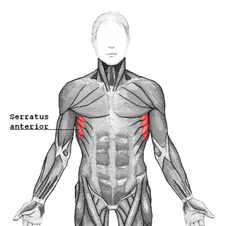
The serratus anterior is a muscle of the chest. It originates at the side of the chest from the upper 8 or 9 ribs; it inserts along the entire length of the anterior aspect of the medial border of the scapula. It is innervated by the long thoracic nerve from the brachial plexus. The serratus anterior acts to pull the scapula forward around the thorax.

<span class="mw-page-title-main">Axillary artery</span> Large blood vessel bringing oxygenated blood to the thorax

In human anatomy, the axillary artery is a large blood vessel that conveys oxygenated blood to the lateral aspect of the thorax, the axilla (armpit) and the upper limb. Its origin is at the lateral margin of the first rib, before which it is called the subclavian artery.

<span class="mw-page-title-main">Subscapular artery</span> Large blood vessel

The subscapular artery, the largest branch of the axillary artery, arises from the third part of the axillary artery at the lower border of the subscapularis muscle, which it follows to the inferior angle of the scapula, where it anastomoses with the lateral thoracic and intercostal arteries, and with the descending branch of the dorsal scapular artery, and ends in the neighboring muscles.

<span class="mw-page-title-main">Occipital artery</span>

The occipital artery is a branch of the external carotid artery that provides arterial supply to the back of the scalp, sternocleidomastoid muscles, and deep muscles of the back and neck.

<span class="mw-page-title-main">Posterior triangle of the neck</span>

The posterior triangle is a region of the neck.

<span class="mw-page-title-main">Thyrocervical trunk</span> Artery of the neck

The thyrocervical trunk is a short artery of the neck. It arises from the subclavian artery, then promptly divides into its branches: the inferior thyroid artery, suprascapular artery, and (sometimes) the transverse cervical artery.

<span class="mw-page-title-main">Circumflex scapular artery</span> Artery of the upper body

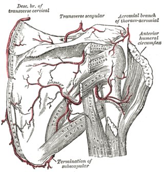
The circumflex scapular artery is a branch of the subscapular artery and part of the scapular anastomoses.

<span class="mw-page-title-main">Suprascapular artery</span> Artery of the neck

The suprascapular artery is a branch of the thyrocervical trunk on the neck.

<span class="mw-page-title-main">Supraspinous fossa</span> Part of the shoulderblade

The supraspinous fossa of the posterior aspect of the scapula is smaller than the infraspinous fossa, concave, smooth, and broader at its vertebral than at its humeral end. Its medial two-thirds give origin to the Supraspinatus.

<span class="mw-page-title-main">Scapular anastomosis</span> Circulatory system around the shoulder blade

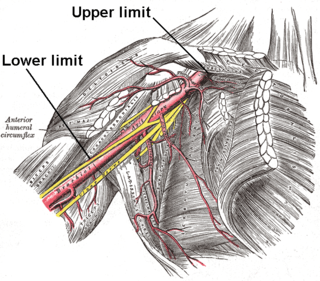
The scapular anastomosis is a system connecting certain subclavian artery and their corresponding axillary artery, forming a circulatory anastomosis around the scapula. It allows blood to flow past the joint in case of occlusion, damage, or pinching of the following scapular arteries:

In anatomy, arterial tree is used to refer to all arteries and/or the branching pattern of the arteries. This article regards the human arterial tree. Starting from the aorta:

<span class="mw-page-title-main">Outline of human anatomy</span> Overview of and topical guide to human anatomy

The following outline is provided as an overview of and topical guide to human anatomy:

<span class="mw-page-title-main">Descending branch of occipital artery</span>

The descending branch of occipital artery, the largest branch of the occipital, descends on the back of the neck, and divides into a superficial and deep portion.

References

  1. Neligan Plastic Surgery, 3rd edition. 2012. pp. Volume 4 Chapter 9 Page 223.
  2. Moore And Agur. Essential Clinical Anatomy (2002) America: Lippincott Williams Publisher. 2nd Ed.
  3. 1 2 "Scapular artery, dorsal". Medcyclopaedia. GE.[ dead link ]
  4. Huelke DF (1962). "The dorsal scapular artery--a proposed term for the artery to the rhomboid muscles" (PDF). Anat. Rec. 142: 57–61. doi:10.1002/ar.1091420109. hdl: 2027.42/49794 . PMID   14449723. S2CID   38155312.
  5. Reiner A, Kasser R (1996). "Relative frequency of a subclavian vs. a transverse cervical origin for the dorsal scapular artery in humans". Anat Rec. 244 (2): 265–8. doi: 10.1002/(SICI)1097-0185(199602)244:2<265::AID-AR14>3.0.CO;2-N . PMID   8808401.

PD-icon.svgThis article incorporates text in the public domain from page 82 of the 20th edition of Gray's Anatomy (1918)