In anatomy, the temporomandibular joints (TMJ) are the two joints connecting the jawbone to the skull. It is a bilateral synovial articulation between the temporal bone of the skull above and the condylar process of mandible below; it is from these bones that its name is derived. The joints are unique in their bilateral function, being connected via the mandible.

In neuroanatomy, the mandibular nerve (V3) is the largest of the three divisions of the trigeminal nerve, the fifth cranial nerve (CN V). Unlike the other divisions of the trigeminal nerve (ophthalmic nerve, maxillary nerve) which contain only afferent fibers, the mandibular nerve contains both afferent and efferent fibers. These nerve fibers innervate structures of the lower jaw and face, such as the tongue, lower lip, and chin. The mandibular nerve also innervates the muscles of mastication.

A pulmonary artery is an artery in the pulmonary circulation that carries deoxygenated blood from the right side of the heart to the lungs. The largest pulmonary artery is the main pulmonary artery or pulmonary trunk from the heart, and the smallest ones are the arterioles, which lead to the capillaries that surround the pulmonary alveoli.

In anatomy, the masseter is one of the muscles of mastication. Found only in mammals, it is particularly powerful in herbivores to facilitate chewing of plant matter. The most obvious muscle of mastication is the masseter muscle, since it is the most superficial and one of the strongest.

The auriculotemporal nerve is a sensory branch of the mandibular nerve (CN V3) that runs with the superficial temporal artery and vein, and provides sensory innervation to parts of the external ear, scalp, and temporomandibular joint. The nerve also conveys post-ganglionic parasympathetic fibres from the otic ganglion to the parotid gland.
The risorius muscle is a highly variable muscle of facial expression. It has numerous and very variable origins, and inserts into the angle of the mouth. It receives motor innervation from branches of facial nerve. It may be absent or asymmetrical in some people. It pulls the angle of the mouth sidewise, such as during smiling.

In human anatomy, the inferior epigastric artery is an artery that arises from the external iliac artery. It is accompanied by the inferior epigastric vein; inferiorly, these two inferior epigastric vessels together travel within the lateral umbilical fold The inferior epigastric artery then traverses the arcuate line of rectus sheath to enter the rectus sheath, then anastomoses with the superior epigastric artery within the rectus sheath.

The pterygoid plexus is a fine venous plexus upon and within the lateral pterygoid muscle. It drains by a short maxillary vein.

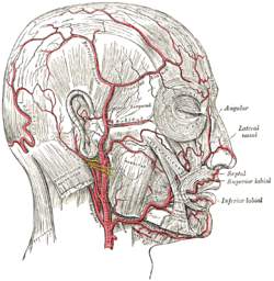
The transverse facial artery is an artery that branches from the superficial temporal artery and runs across the face.

The maxillary artery supplies deep structures of the face. It branches from the external carotid artery just deep to the neck of the mandible.

The infratemporal fossa is an irregularly shaped cavity that is a part of the skull. It is situated below and medial to the zygomatic arch. It is not fully enclosed by bone in all directions. It contains superficial muscles, including the lower part of the temporalis muscle, the lateral pterygoid muscle, and the medial pterygoid muscle. It also contains important blood vessels such as the middle meningeal artery, the pterygoid plexus, and the retromandibular vein, and nerves such as the mandibular nerve (CN V3) and its branches.
The masseteric nerve is a nerve of the face. It is a branch of the mandibular nerve (CN V3). It passes through the mandibular notch to reach masseter muscle. It provides motor innervation the masseter muscle, and sensory innervation to the temporomandibular joint.

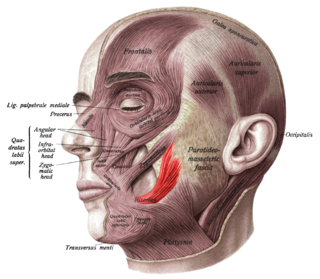
The masseteric fascia and parotideomasseteric fascia are fascias of the head varyingly described depending upon the source consulted. They may or may not be described as one and the same structure.
Masseteric is an adjective meaning "of or pertaining to the Masseter muscle", such as:
The mandibular notch, also known as the sigmoid notch, is a groove in the ramus of the mandible. It is the gap between the coronoid process anteriorly and the condyloid process posteriorly.
In anatomy, arterial tree is used to refer to all arteries and/or the branching pattern of the arteries. This article regards the human arterial tree. Starting from the aorta:

The intercostal arteries are a group of arteries passing within an intercostal space. There are 9 anterior and 11 posterior intercostal arteries on each side of the body. The anterior intercostal arteries are branches of the internal thoracic artery and its terminal branch – the musculophrenic artery. The posterior intercostal arteries are branches of the supreme intercostal artery and thoracic aorta.

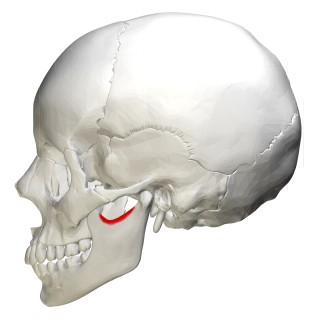
The angle of the mandible is located at the posterior border at the junction of the lower border of the ramus of the mandible.

The following outline is provided as an overview of and topical guide to human anatomy:
The parotid fascia is a tough fascia enclosing the parotid gland. It has a superficial layer and a deep layer.