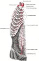
In human anatomy, the subclavian arteries are paired major arteries of the upper thorax, below the clavicle. They receive blood from the aortic arch. The left subclavian artery supplies blood to the left arm and the right subclavian artery supplies blood to the right arm, with some branches supplying the head and thorax. On the left side of the body, the subclavian comes directly off the aortic arch, while on the right side it arises from the relatively short brachiocephalic artery when it bifurcates into the subclavian and the right common carotid artery.

The popliteal artery is a deeply placed continuation of the femoral artery opening in the distal portion of the adductor magnus muscle. It courses through the popliteal fossa and ends at the lower border of the popliteus muscle, where it branches into the anterior and posterior tibial arteries.

The vertebral arteries are major arteries of the neck. Typically, the vertebral arteries originate from the subclavian arteries. Each vessel courses superiorly along each side of the neck, merging within the skull to form the single, midline basilar artery. As the supplying component of the vertebrobasilar vascular system, the vertebral arteries supply blood to the upper spinal cord, brainstem, cerebellum, and posterior part of brain.

The auriculotemporal nerve is a sensory branch of the mandibular nerve (CN V3) that runs with the superficial temporal artery and vein, and provides sensory innervation to parts of the external ear, scalp, and temporomandibular joint. The nerve also conveys post-ganglionic parasympathetic fibres from the otic ganglion to the parotid gland.
In anatomy, the left and right common carotid arteries (carotids) are arteries that supply the head and neck with oxygenated blood; they divide in the neck to form the external and internal carotid arteries.

The sternohyoid muscle is a bilaterally paired, long, thin, narrow strap muscle of the anterior neck. It is one of the infrahyoid muscles. It is innervated by the ansa cervicalis. It acts to depress the hyoid bone.

In human anatomy, the inferior epigastric artery is an artery that arises from the external iliac artery. It is accompanied by the inferior epigastric vein; inferiorly, these two inferior epigastric vessels together travel within the lateral umbilical fold The inferior epigastric artery then traverses the arcuate line of rectus sheath to enter the rectus sheath, then anastomoses with the superior epigastric artery within the rectus sheath.

In human anatomy, the superior epigastric artery is a terminal branch of the internal thoracic artery that provides arterial supply to the abdominal wall, and upper rectus abdominis muscle. It enters the rectus sheath to descend upon the inner surface of the rectus abdominis muscle. It ends by anastomosing with the inferior epigastric artery.

The posterior triangle is a region of the neck.

The inferior gluteal artery is a terminal branch of the anterior trunk of the internal iliac artery. It exits the pelvis through the greater sciatic foramen. It is distributed chiefly to the buttock and the back of the thigh.

The transverse cervical artery is an artery in the neck and a branch of the thyrocervical trunk, running at a higher level than the suprascapular artery.
The inferior thyroid artery is an artery in the neck. It arises from the thyrocervical trunk and passes upward, in front of the vertebral artery and longus colli muscle. It then turns medially behind the carotid sheath and its contents, and also behind the sympathetic trunk, the middle cervical ganglion resting upon the vessel.

The lateral circumflex femoral artery is an artery in the upper thigh. It is usually a branch of the profunda femoris artery, and produces three branches. It is mostly distributed to the muscles of the lateral thigh, supplying arterial blood to muscles of the knee extensor group.
The suprascapular artery is a branch of the thyrocervical trunk on the neck.

The scapular anastomosis is a system connecting certain subclavian artery and their corresponding axillary artery, forming a circulatory anastomosis around the scapula. It allows blood to flow past the joint in case of occlusion, damage, or pinching of the following scapular arteries:

The costocervical trunk arises from the upper and back part of the second part of subclavian artery, behind the scalenus anterior on the right side, and medial to that muscle on the left side.

The subclavian triangle, the smaller division of the posterior triangle, is bounded, above, by the inferior belly of the omohyoideus; below, by the clavicle; its base is formed by the posterior border of the sternocleidomastoideus.

The inferior cervical ganglion is one of the three cervical sympathetic ganglia. It situated between the base of the transverse process of the last cervical vertebra and the neck of the first rib, on the medial side of the costocervical artery.

The prevertebral fascia is the layer of deep cervical fascia that surrounds the vertebral column. It is the deepest layer of deep cervical fascia.

The following outline is provided as an overview of and topical guide to human anatomy: