Related Research Articles

The sella turcica is a saddle-shaped depression in the body of the sphenoid bone of the human skull and of the skulls of other hominids including chimpanzees, gorillas and orangutans. It serves as a cephalometric landmark. The pituitary gland or hypophysis is located within the most inferior aspect of the sella turcica, the hypophyseal fossa.
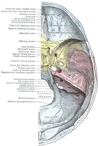
The foramen lacerum is a triangular hole in the base of skull. It is located between the sphenoid bone, the apex of the petrous part of the temporal bone, and the basilar part of the occipital bone.

The celiacartery, also known as the celiac trunk or truncus coeliacus, is the first major branch of the abdominal aorta. It is about 1.25 cm in length. Branching from the aorta at thoracic vertebra 12 (T12) in humans, it is one of three anterior/ midline branches of the abdominal aorta.

The ophthalmic artery (OA) is an artery of the head. It is the first branch of the internal carotid artery distal to the cavernous sinus. Branches of the ophthalmic artery supply all the structures in the orbit around the eye, as well as some structures in the nose, face, and meninges. Occlusion of the ophthalmic artery or its branches can produce sight-threatening conditions.

The facial artery is a branch of the external carotid artery that supplies structures of the superficial face.

The cavernous sinus within the human head is one of the dural venous sinuses creating a cavity called the lateral sellar compartment bordered by the temporal bone of the skull and the sphenoid bone, lateral to the sella turcica.

The carotid sheath is a condensation of the deep cervical fascia enveloping multiple vital neurovascular structures of the neck, including the common and internal carotid arteries, the internal jugular vein, the vagus nerve, and ansa cervicalis. The carotid sheath helps protects the structures contained therein.

In human anatomy, the inferior epigastric artery is an artery that arises from the external iliac artery. It is accompanied by the inferior epigastric vein; inferiorly, these two inferior epigastric vessels together travel within the lateral umbilical fold The inferior epigastric artery then traverses the arcuate line of rectus sheath to enter the rectus sheath, then anastomoses with the superior epigastric artery within the rectus sheath.

The lingual artery arises from the external carotid artery between the superior thyroid artery and facial artery. It can be located easily in the tongue.

The sphenoid sinus is a paired paranasal sinus occurring within the body of the sphenoid bone. It represents one pair of the four paired paranasal sinuses. The pair of sphenoid sinuses are separated in the middle by a septum of sphenoid sinuses. Each sphenoid sinus communicates with the nasal cavity via the opening of sphenoidal sinus. The two sphenoid sinuses vary in size and shape, and are usually asymmetrical.

The iliolumbar artery is the first branch of the posterior trunk of the internal iliac artery.

The lateral sacral arteries is an artery in the pelvis that arises from the posterior division of the internal iliac artery. It later splits into two smaller branches, a superior and an inferior.

The inferior thyroid artery is an artery in the neck. It arises from the thyrocervical trunk and passes upward, in front of the vertebral artery and longus colli muscle. It then turns medially behind the carotid sheath and its contents, and also behind the sympathetic trunk, the middle cervical ganglion resting upon the vessel.

The perineal nerve is a nerve of the pelvis. It arises from the pudendal nerve in the pudendal canal. It gives superficial branches to the skin, and a deep branch to muscles. It supplies the skin and muscles of the perineum. Its latency is tested with electrodes.

The carotid canal is a passage in the petrous part of the temporal bone of the skull through which the internal carotid artery and its internal carotid (nervous) plexus pass from the neck into the cranial cavity.

The retromandibular vein is a major vein of the face. It is formed within the parotid gland by the confluence of the maxillary vein, and superficial temporal vein. It descends in the gland and splits into two branches upon emerging from the gland. Its anterior branch then joins the (anterior) facial vein forming the common facial vein, while its posterior branch joins the posterior auricular vein forming the external jugular vein.

The infratemporal fossa is an irregularly shaped cavity that is a part of the skull. It is situated below and medial to the zygomatic arch. It is not fully enclosed by bone in all directions. It contains superficial muscles, including the lower part of the temporalis muscle, the lateral pterygoid muscle, and the medial pterygoid muscle. It also contains important blood vessels such as the middle meningeal artery, the pterygoid plexus, and the retromandibular vein, and nerves such as the mandibular nerve (CN V3) and its branches.
The posterior ethmoidal nerve is a nerve of the head. It is a branch of the nasociliary nerve (itself a branch of the ophthalmic nerve (CN V1)). It provides sensory innervation to the sphenoid sinus and ethmoid sinus, and part of the dura mater in the anterior cranial fossa.

The anterior clinoid process is a posterior projection of the sphenoid bone at the junction of the medial end of either lesser wing of sphenoid bone with the body of sphenoid bone. The bilateral processes flank the sella turcica anteriorly.

The inferior suprarenal artery is a paired artery that supplies the adrenal gland. It usually originates at the trunk of the renal artery before its terminal division, but with many common variations. It supplies the adrenal gland parenchyma, the ureter, and the surrounding cellular tissue and muscles.
References
- ↑ Gibo H, Hokama M, Kyoshima K, Kobayashi S (1993). "Arteries to the pituitary". Nippon Rinsho. 51 (10): 2550–4. PMID 8254920.
- ↑ Marieb, Elaine (2014). Anatomy & physiology. Glenview, IL: Pearson Education, Inc. ISBN 978-0321861580.
- 1 2 3 4 5 Seker, Askin; Martins, Carolina; Rhoton Jr., Albert L. (2010). "2 - Meningeal Anatomy". Meningiomas. Saunders. pp. 11–51. doi:10.1016/B978-1-4160-5654-6.00002-7. ISBN 978-1-4160-5654-6.
- ↑ Maynard, Robert Lewis; Downes, Noel (2019). "16 - Endocrine Glands". Anatomy and Histology of the Laboratory Rat in Toxicology and Biomedical Research. Academic Press. pp. 185–196. doi:10.1016/B978-0-12-811837-5.00016-2. ISBN 978-0-12-811837-5. S2CID 239275973.
- ↑ Johnson, Mark (2010). "11 - Endocrinology". Basic Science in Obstetrics and Gynaecology (4th ed.). Churchill Livingstone. pp. 231–257. doi:10.1016/B978-0-443-10281-3.00015-4. ISBN 978-0-443-10281-3.
- ↑ Frenette, Eric; Lui, Alben; Cao, Michelle (2012). "1 - Neurohormones and Sleep". Vitamins & Hormones. Vol. 89. Elsevier. pp. 1–17. doi:10.1016/B978-0-12-394623-2.00001-9. ISBN 978-0-12-394623-2. ISSN 0083-6729. PMID 22640605.