The external jugular vein receives the greater part of the blood from the exterior of the cranium and the deep parts of the face, being formed by the junction of the posterior division of the retromandibular vein with the posterior auricular vein.

The inferior petrosal sinuses are two small sinuses situated on the inferior border of the petrous part of the temporal bone, one on each side. Each inferior petrosal sinus drains the cavernous sinus into the internal jugular vein.

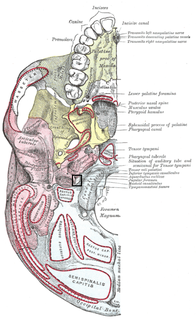
The jugular fossa is a deep depression in the inferior part of the base of the skull. More specifically, it is located in the temporal bone, posterior to the carotid canal and the aquæductus cochleæ. It is of variable depth and size in different skulls; it lodges the bulb of the internal jugular vein.

The anterior jugular vein is a vein in the neck. It begins near the hyoid bone by the confluence of several superficial veins from the submandibular region.
The Facial vein usually unites with the anterior branch of the Retromandibular vein to form the Common Facial Vein, which crosses the external carotid artery and enters the internal jugular vein at a variable point below the hyoid bone.
The retromandibular vein, formed by the union of the superficial temporal and maxillary veins, descends in the substance of the parotid gland, superficial to the external carotid artery but beneath the facial nerve, between the ramus of the mandible and the sternocleidomastoideus muscle.

The lateral parts of the occipital bone are situated at the sides of the foramen magnum; on their under surfaces are the condyles for articulation with the superior facets of the atlas.

The superior ganglion of the vagus nerve or jugular ganglion is a well-marked ganglionic enlargement of the vagus nerve. It is located in the middle part of the jugular foramen. It contains afferent somatosensory neuronal cell bodies that provide sensory information from the external auditory meatus, cranial meninges, and the external surface of the tympanic membrane. Their central fibers synapse in the Trigeminal nerve nuclei.
In the lateral part of the occipital bone, extending lateralward from the posterior half of the condyle is a quadrilateral or triangular plate of bone, the jugular process, excavated in front by the jugular notch, which, in the articulated skull, forms the posterior part of the jugular foramen.

Medial to the opening for the carotid canal and close to its posterior border, in front of the jugular fossa, is a triangular depression; at the apex of this is a small opening, the aquaeductus cochleae, which lodges a tubular prolongation of the dura mater establishing a communication between the perilymphatic space and the subarachnoid space, and transmits a vein from the cochlea to join the internal jugular vein.

The right lymphatic duct, about 1.25 cm. in length, courses along the medial border of the Scalenus anterior at the root of the neck. The right lymphatic duct forms various combinations with the right subclavian vein and right internal jugular vein. A right lymphatic duct that enters directly into the junction of the internal jugular and subclavian veins is uncommon. The discovery of this structure has been credited to Niels Stensen.

An apical group of six to twelve glands is situated partly posterior to the upper portion of the Pectoralis minor and partly above the upper border of this muscle.

The deep cervical lymph nodes are a group of cervical lymph nodes found near the internal jugular vein.

The jugular trunk is a lymphatic vessel in the neck. It is formed by vessels that emerge from the superior deep cervical lymph nodes and unite to efferents of the inferior deep cervical lymph nodes.

The superior deep cervical lymph nodes lie under the sternocleidomastoid muscle in close relation with the accessory nerve and the internal jugular vein.
Just above the sternum the two anterior jugular veins communicate by a transverse trunk, the jugular venous arch, which receive tributaries from the inferior thyroid veins; each also communicates with the internal jugular.
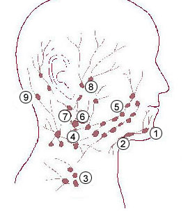
The superficial cervical lymph nodes are lymph nodes that lie near the surface of the neck.
The efferent vessels of the subclavicular group unite to form the subclavian trunk, which opens either directly into the junction of the internal jugular and subclavian veins or into the jugular lymphatic trunk; on the left side it may end in the thoracic duct.
The public domain consists of all the creative works to which no exclusive intellectual property rights apply. Those rights may have expired, been forfeited, expressly waived, or may be inapplicable.

Gray's Anatomy is an English language textbook of human anatomy originally written by Henry Gray and illustrated by Henry Vandyke Carter. Earlier editions were called Anatomy: Descriptive and Surgical, Anatomy of the Human Body and Gray's Anatomy: Descriptive and Applied, but the book's name is commonly shortened to, and later editions are titled, Gray's Anatomy. The book is widely regarded as an extremely influential work on the subject, and has continued to be revised and republished from its initial publication in 1858 to the present day. The latest edition of the book, the 41st, was published in September 2015.