Cerebral circulation

Last updated

Cerebral circulation
Cerebral vascular territories.jpg
Areas of the brain are supplied by different arteries. The major systems are divided into an anterior circulation (the anterior cerebral artery and middle cerebral artery) and a posterior circulation.
Gray488 blue.gif
Schematic of veins and venous spaces that drain deoxygenated blood from the brain
Identifiers
MeSH D002560
Anatomical terminology

Cerebral circulation is the movement of blood through a network of cerebral arteries and veins supplying the brain. The rate of cerebral blood flow in an adult human is typically 750 milliliters per minute, or about 15% of cardiac output. Arteries deliver oxygenated blood, glucose and other nutrients to the brain. Veins carry "used or spent" blood back to the heart, to remove carbon dioxide, lactic acid, and other metabolic products. The neurovascular unit regulates cerebral blood flow so that activated neurons can be supplied with energy in the right amount and at the right time. [1] Because the brain would quickly suffer damage from any stoppage in blood supply, the cerebral circulatory system has safeguards including autoregulation of the blood vessels. The failure of these safeguards may result in a stroke. The volume of blood in circulation is called the cerebral blood flow. Sudden intense accelerations change the gravitational forces perceived by bodies and can severely impair cerebral circulation and normal functions to the point of becoming serious life-threatening conditions.

Contents

The following description is based on idealized human cerebral circulation. The pattern of circulation and its nomenclature vary between organisms.

Anatomy

Cerebrovascular system Cerebrovascular System.png
Cerebrovascular system

Blood supply

Cerebral vascular territories.jpg
Cerebral vascular territories midline.jpg
Cortical areas and their arterial blood supply

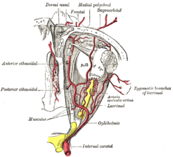
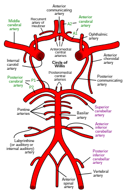
Blood supply to the brain is normally divided into anterior and posterior segments, relating to the different arteries that supply the brain. The two main pairs of arteries are the internal carotid arteries (supply the anterior brain) and vertebral arteries (supplying the brainstem and posterior brain). [2] The anterior and posterior cerebral circulations are interconnected via bilateral posterior communicating arteries. They are part of the circle of Willis, which provides backup circulation to the brain. In case one of the supply arteries is occluded, the circle of Willis provides interconnections between the anterior and the posterior cerebral circulation along the floor of the cerebral vault, providing blood to tissues that would otherwise become ischemic. [3]

Anterior cerebral circulation

The ophthalmic artery and its branches Gray514.png
The ophthalmic artery and its branches

The anterior cerebral circulation is the blood supply to the anterior portion of the brain including eyes. It is supplied by the following arteries:

Posterior cerebral circulation

The anterior and posterior circulations meet at the circle of Willis, pictured here, which rests at the top of the brainstem. Inferior view. Circle of Willis en.svg
The anterior and posterior circulations meet at the circle of Willis, pictured here, which rests at the top of the brainstem. Inferior view.

The posterior cerebral circulation is the blood supply to the posterior portion of the brain, including the occipital lobes, cerebellum and brainstem. It is supplied by the following arteries:

Venous drainage

The venous drainage of the cerebrum can be separated into two subdivisions: superficial and deep.

The superficial system

The superficial system is composed of dural venous sinuses, sinuses (channels) within the dura mater. The dural sinuses are therefore located on the surface of the cerebrum. The most prominent of these sinuses is the superior sagittal sinus which is located in the sagittal plane under the midline of the cerebral vault, posteriorly and inferiorly to the confluence of sinuses, where the superficial drainage joins with the sinus that primarily drains the deep venous system. From here, two transverse sinuses bifurcate and travel laterally and inferiorly in an S-shaped curve that forms the sigmoid sinuses which go on to form the two jugular veins. In the neck, the jugular veins parallel the upward course of the carotid arteries and drain blood into the superior vena cava. The veins puncture the relevant dural sinus, piercing the arachnoid and dura mater as bridging veins that drain their contents into the sinus. [5]

The deep venous system

The deep venous system is primarily composed of traditional veins inside the deep structures of the brain, which join behind the midbrain to form the great cerebral vein (vein of Galen). This vein merges with the inferior sagittal sinus to form the straight sinus which then joins the superficial venous system mentioned above at the confluence of sinuses.

Maturation of cerebral blood vessels

The maturation of blood vessels in the brain is a critical process that occurs postnatally. [6] It involves the acquisition of key barrier and contractile properties essential for brain function. During the early postnatal phase, endothelial cells (ECs) and vascular smooth muscle cells (VSMCs) undergo significant molecular and functional changes.

Endothelial cells begin to express P-glycoprotein, a crucial efflux transporter that helps protect the brain by expelling harmful substances. [7] This efflux capacity is progressively acquired and becomes fully functional by the postnatal period​​. Additionally, VSMCs, which initially populate the arterial network, start to express contractile proteins such as smooth muscle actin (SMA) and myosin-11, transforming VSMCs into contractile cells capable of regulating blood vessel tone and cerebral blood flow​​.

The expression of Myh11 in VSMCs acts as a developmental switch, with significant upregulation occurring from birth to the age of 2 to 5 years. [6] This is a critical period needed for the establishment of vessel contractility and the overall functionality of the cerebral circulation.

Physiology

Dural venous sinuses bordered by hard meninges (shown in blue) direct blood outflow from cerebral veins to the internal jugular vein at the base of skull. Sobo 1909 589.png
Dural venous sinuses bordered by hard meninges (shown in blue) direct blood outflow from cerebral veins to the internal jugular vein at the base of skull.

Cerebral blood flow (CBF) is the blood supply to the brain in a given period of time. [8] In an adult, CBF is typically 750 millilitres per minute or 15.8 ± 5.7% of the cardiac output. [9] This equates to an average perfusion of 50 to 54 millilitres of blood per 100 grams of brain tissue per minute. [10] [11] [12]

The radio index of cerebral blood flow/cardiac output (CCRI) decreases by 1.3% per decade, even though cardiac output remains unchanged. [9]  Across the adult lifespan, women have a higher CCRI than men. [9] CBF is inversely associated with body mass index. [9]

CBF is tightly regulated to meet the brain's metabolic demands. [10] [13] Too much blood (a clinical condition of a normal homeostatic response of hyperemia) [1] can raise intracranial pressure (ICP), which can compress and damage delicate brain tissue. Too little blood flow (ischemia) results if blood flow to the brain is below 18 to 20 ml per 100 g per minute, and tissue death occurs if flow dips below 8 to 10 ml per 100 g per minute. In brain tissue, a biochemical cascade known as the ischemic cascade is triggered when the tissue becomes ischemic, potentially resulting in damage to and the death of brain cells. Medical professionals must take steps to maintain proper CBF in patients who have conditions like shock, stroke, cerebral edema, and traumatic brain injury.

Cerebral blood flow is determined by a number of factors, such as viscosity of blood, how dilated blood vessels are, and the net pressure of the flow of blood into the brain, known as cerebral perfusion pressure, which is determined by the body's blood pressure. Cerebral perfusion pressure (CPP) is defined as the mean arterial pressure (MAP) minus the intracranial pressure (ICP). In normal individuals, it should be above 50 mm Hg. Intracranial pressure should not be above 15 mm Hg (ICP of 20 mm Hg is considered as intracranial hypertension). [14] Cerebral blood vessels are able to change the flow of blood through them by altering their diameters in a process called cerebral autoregulation; they constrict when systemic blood pressure is raised and dilate when it is lowered. [15] Arterioles also constrict and dilate in response to different chemical concentrations. For example, they dilate in response to higher levels of carbon dioxide in the blood and constrict in response to lower levels of carbon dioxide. [15]

For example, assuming a person with an arterial partial pressure of carbon dioxide (PaCO2) of 40 mmHg (normal range of 38–42 mmHg) [16] and a CBF of 50 ml per 100g per min. If the PaCO2 dips to 30 mmHg, this represents a 10 mmHg decrease from the initial value of PaCO2. Consequently, the CBF decreases by 1ml per 100g per min for each 1mmHg decrease in PaCO2, resulting in a new CBF of 40ml per 100g of brain tissue per minute. In fact, for each 1 mmHg increase or decrease in PaCO2, between the range of 20–60 mmHg, there is a corresponding CBF change in the same direction of approximately 1–2 ml/100g/min, or 2–5% of the CBF value. [17] This is why small alterations in respiration pattern can cause significant changes in global CBF, specially through PaCO2 variations. [17]

CBF is equal to the cerebral perfusion pressure (CPP) divided by the cerebrovascular resistance (CVR): [18]

CBF = CPP / CVR

Control of CBF is considered in terms of the factors affecting CPP and the factors affecting CVR. CVR is controlled by four major mechanisms:

  1. Metabolic control (or 'metabolic autoregulation')
  2. Pressure autoregulation
  3. Chemical control (by arterial pCO2 and pO2)
  4. Neural control

Role of intracranial pressure

Increased intracranial pressure (ICP) causes decreased blood perfusion of brain cells by mainly two mechanisms:

Cerebral perfusion pressure

Cerebral perfusion pressure is the net pressure gradient causing cerebral blood flow to the brain (brain perfusion). It must be maintained within narrow limits; too little pressure could cause brain tissue to become ischemic (having inadequate blood flow), and too much could raise intracranial pressure.

Imaging

Arterial spin labeling (ASL), phase contrast magnetic resonance imaging (PC-MRI), and positron emission tomography (PET) are neuroimaging techniques that can be used to measure CBF. ASL and PET can also be used to measure regional CBF (rCBF) within a specific brain region. rCBF at one location can be measured over time by thermal diffusion [19]

Related Research Articles

<span class="mw-page-title-main">Vein</span> Blood vessels that carry blood towards the heart

Veins are blood vessels in the circulatory system of humans and most other animals that carry blood towards the heart. Most veins carry deoxygenated blood from the tissues back to the heart; exceptions are those of the pulmonary and fetal circulations which carry oxygenated blood to the heart. In the systemic circulation, arteries carry oxygenated blood away from the heart, and veins return deoxygenated blood to the heart, in the deep veins.

<span class="mw-page-title-main">Coronary circulation</span> Circulation of blood in the blood vessels of the heart muscle (myocardium)

Coronary circulation is the circulation of blood in the arteries and veins that supply the heart muscle (myocardium). Coronary arteries supply oxygenated blood to the heart muscle. Cardiac veins then drain away the blood after it has been deoxygenated. Because the rest of the body, and most especially the brain, needs a steady supply of oxygenated blood that is free of all but the slightest interruptions, the heart is required to function continuously. Therefore its circulation is of major importance not only to its own tissues but to the entire body and even the level of consciousness of the brain from moment to moment. Interruptions of coronary circulation quickly cause heart attacks, in which the heart muscle is damaged by oxygen starvation. Such interruptions are usually caused by coronary ischemia linked to coronary artery disease, and sometimes to embolism from other causes like obstruction in blood flow through vessels.

<span class="mw-page-title-main">Circle of Willis</span> Circulatory anastomosis that supplies blood to the brain and surrounding structures

The circle of Willis is a circulatory anastomosis that supplies blood to the brain and surrounding structures in reptiles, birds and mammals, including humans. It is named after Thomas Willis (1621–1675), an English physician.

<span class="mw-page-title-main">Cerebral edema</span> Excess accumulation of fluid (edema) in the intracellular or extracellular spaces of the brain

Cerebral edema is excess accumulation of fluid (edema) in the intracellular or extracellular spaces of the brain. This typically causes impaired nerve function, increased pressure within the skull, and can eventually lead to direct compression of brain tissue and blood vessels. Symptoms vary based on the location and extent of edema and generally include headaches, nausea, vomiting, seizures, drowsiness, visual disturbances, dizziness, and in severe cases, death.

<span class="mw-page-title-main">Intracranial pressure</span> Pressure exerted by fluids inside the skull and on the brain

Intracranial pressure (ICP) is the pressure exerted by fluids such as cerebrospinal fluid (CSF) inside the skull and on the brain tissue. ICP is measured in millimeters of mercury (mmHg) and at rest, is normally 7–15 mmHg for a supine adult. This equals to 9–20 cmH2O, which is a common scale used in lumbar punctures. The body has various mechanisms by which it keeps the ICP stable, with CSF pressures varying by about 1 mmHg in normal adults through shifts in production and absorption of CSF.

<span class="mw-page-title-main">Internal carotid artery</span> Artery supplying the brain

The internal carotid artery is an artery in the neck which supplies the anterior and middle cerebral circulation.

<span class="mw-page-title-main">Dura mater</span> Outermost layer of the protective tissues around the central nervous system (meninges)

In neuroanatomy, dura mater is a thick membrane made of dense irregular connective tissue that surrounds the brain and spinal cord. It is the outermost of the three layers of membrane called the meninges that protect the central nervous system. The other two meningeal layers are the arachnoid mater and the pia mater. It envelops the arachnoid mater, which is responsible for keeping in the cerebrospinal fluid. It is derived primarily from the neural crest cell population, with postnatal contributions of the paraxial mesoderm.

<span class="mw-page-title-main">Intracranial hemorrhage</span> Hemorrhage, or bleeding, within the skull

Intracranial hemorrhage (ICH), also known as intracranial bleed, is bleeding within the skull. Subtypes are intracerebral bleeds, subarachnoid bleeds, epidural bleeds, and subdural bleeds.

<span class="mw-page-title-main">Cerebral angiography</span> Angiography that produces images of blood vessels in and around the brain

Cerebral angiography is a form of angiography which provides images of blood vessels in and around the brain, thereby allowing detection of abnormalities such as arteriovenous malformations and aneurysms. It was pioneered in 1927 by the Portuguese neurologist Egas Moniz at the University of Lisbon, who also helped develop thorotrast for use in the procedure.

Cushing reflex is a physiological nervous system response to increased intracranial pressure (ICP) that results in Cushing's triad of increased blood pressure, irregular breathing, and bradycardia. It is usually seen in the terminal stages of acute head injury and may indicate imminent brain herniation. It can also be seen after the intravenous administration of epinephrine and similar drugs. It was first described in detail by American neurosurgeon Harvey Cushing in 1901.

<span class="mw-page-title-main">Internal jugular vein</span> Blood vessel that drains the head

The internal jugular vein is a paired jugular vein that collects blood from the brain and the superficial parts of the face and neck. This vein runs in the carotid sheath with the common carotid artery and vagus nerve.

The emissary veins connect the extracranial venous system with the intracranial venous sinuses. They connect the veins outside the cranium to the venous sinuses inside the cranium. They drain from the scalp, through the skull, into the larger meningeal veins and dural venous sinuses. They may also connect to diploic veins within the skull.

<span class="mw-page-title-main">Intraparenchymal hemorrhage</span> Bleeding within parenchymal tissue of the brain

Intraparenchymal hemorrhage (IPH) is one form of intracerebral bleeding in which there is bleeding within brain parenchyma. The other form is intraventricular hemorrhage (IVH).

Cerebral perfusion pressure, or CPP, is the net pressure gradient causing cerebral blood flow to the brain. It must be maintained within narrow limits because too little pressure could cause brain tissue to become ischemic, and too much could raise intracranial pressure (ICP).

<span class="mw-page-title-main">Posterior communicating artery</span> Arteries at the base of the brain that form part of the circle of Willis

In human anatomy, the left and right posterior communicating arteries are small arteries at the base of the brain that form part of the circle of Willis.

<span class="mw-page-title-main">Cerebral infarction</span> Stroke resulting from lack of blood flow

Cerebral infarction, also known as an ischemic stroke, is the pathologic process that results in an area of necrotic tissue in the brain. In mid to high income countries, a stroke is the main reason for disability among people and the 2nd cause of death. It is caused by disrupted blood supply (ischemia) and restricted oxygen supply (hypoxia). This is most commonly due to a thrombotic occlusion, or an embolic occlusion of major vessels which leads to a cerebral infarct. In response to ischemia, the brain degenerates by the process of liquefactive necrosis.

<span class="mw-page-title-main">Dural arteriovenous fistula</span> Medical condition

A dural arteriovenous fistula (DAVF) or malformation is an abnormal direct connection (fistula) between a meningeal artery and a meningeal vein or dural venous sinus.

A circulatory anastomosis is a connection between two blood vessels, such as between arteries, between veins or between an artery and a vein. Anastomoses between arteries and between veins result in a multitude of arteries and veins, respectively, serving the same volume of tissue. Such anastomoses occur normally in the body in the circulatory system, serving as back-up routes in a collateral circulation that allow blood to flow if one link is blocked or otherwise compromised, but may also occur pathologically.

<span class="mw-page-title-main">Leptomeningeal collateral circulation</span>

The leptomeningeal collateral circulation is a network of small blood vessels in the brain that connects branches of the middle, anterior and posterior cerebral arteries, with variation in its precise anatomy between individuals. During a stroke, leptomeningeal collateral vessels allow limited blood flow when other, larger blood vessels provide inadequate blood supply to a part of the brain.

<span class="mw-page-title-main">Spaceflight associated neuro-ocular syndrome</span>

Spaceflight associated neuro-ocular syndrome (SANS), previously called spaceflight-induced visual impairment, is hypothesized to be a result of increased intracranial pressure (ICP), although, experiments directly measuring ICP in parabolic flight have shown ICP to be in normal physiological ranges during acute weightless exposure. The study of visual changes and ICP in astronauts on long-duration flights is a relatively recent topic of interest to space medicine professionals. Although reported signs and symptoms have not appeared to be severe enough to cause blindness in the near term, long term consequences of chronically elevated intracranial pressure are unknown.

References

  1. 1 2 Muoio V, Persson PB, Sendeski MM (April 2014). "The neurovascular unit - concept review". Acta Physiologica. 210 (4): 790–798. doi: 10.1111/apha.12250 . PMID   24629161. S2CID   25274791.
  2. Cipolla MJ (2009). "Chapter 2: Anatomy and Ultrastructure.". The Cerebral Circulation. San Rafael (CA): Morgan & Claypool Life Sciences.
  3. Chandra A, Li WA, Stone CR, Geng X, Ding Y (2017-07-17). "The cerebral circulation and cerebrovascular disease I: Anatomy". Brain Circulation. 3 (2): 45–56. doi: 10.4103/bc.bc_10_17 . PMC   6126264 . PMID   30276305.
  4. Oiseth S, Jones L, Maza E (eds.). "Carotid Arterial System". The Lecturio Medical Concept Library. Retrieved 2021-06-22.
  5. Hufnagle JJ, Tadi P (2022). "Neuroanatomy, Brain Veins". StatPearls. StatPearls Publishing. PMID   31536212 . Retrieved 28 February 2023.
  6. 1 2 Slaoui L, Gilbert A, Rancillac A, Delaunay-Piednoir B, Chagnot A, Gerard Q, et al. (March 2023). "In mice and humans, brain microvascular contractility matures postnatally". Brain Structure & Function. 228 (2): 475–492. doi:10.1007/s00429-022-02592-w. PMID   36380034.
  7. Löscher W, Potschka H (January 2005). "Blood-brain barrier active efflux transporters: ATP-binding cassette gene family". NeuroRx. 2 (1): 86–98. doi:10.1602/neurorx.2.1.86. PMC   539326 . PMID   15717060.
  8. Tolias C, Sgouros S (2006). "Initial Evaluation and Management of CNS Injury". Emedicine.com. Archived from the original on March 2, 2007. Retrieved January 4, 2007.
  9. 1 2 3 4 Xing CY, Tarumi T, Liu J, Zhang Y, Turner M, Riley J, et al. (August 2017). "Distribution of cardiac output to the brain across the adult lifespan". Journal of Cerebral Blood Flow and Metabolism. 37 (8): 2848–2856. doi:10.1177/0271678X16676826. PMC   5536794 . PMID   27789785.
  10. 1 2 "Overview of Adult Traumatic Brain Injuries" (PDF). Orlando Regional Healthcare, Education and Development. 2004. Archived from the original (PDF) on 27 February 2008. Retrieved 16 January 2008.
  11. Shepherd S (2004). "Head Trauma". Emedicine.com. Retrieved 4 January 2007.
  12. Walters FJ (1998). "Intracranial Pressure and Cerebral Blood Flow". Physiology (8). World Federation of Societies of Anaesthesiologists: 4. Archived from the original on 14 May 2011. Retrieved 4 January 2007.
  13. Singh J, Stock A (2006). "Head Trauma". Emedicine.com. Retrieved 4 January 2007.
  14. Mattle H, Mumenthaler M, Taub E (2016-12-14). Fundamentals of Neurology. Thieme. p. 129. ISBN   978-3-13-136452-4.
  15. 1 2 Kandel ER, Schwartz JH, Jessell TM (2000). Principles of Neural Science (4th ed.). New York: McGraw-Hill. p. 1305.
  16. Hadjiliadis D, Zieve D, Ogilvie I (6 June 2015). "Blood Gases". Medline Plus.
  17. 1 2 Giardino ND, Friedman SD, Dager SR (2007). "Anxiety, respiration, and cerebral blood flow: implications for functional brain imaging". Comprehensive Psychiatry. 48 (2): 103–112. doi:10.1016/j.comppsych.2006.11.001. PMC   1820771 . PMID   17292699.
  18. "Cerebral Blood Flow (CBF)". Anaesthesia UK. 2007. Archived from the original on 18 September 2010. Retrieved 16 October 2007.
  19. Vajkoczy P, Roth H, Horn P, Lucke T, Thomé C, Hubner U, et al. (August 2000). "Continuous monitoring of regional cerebral blood flow: experimental and clinical validation of a novel thermal diffusion microprobe". Journal of Neurosurgery. 93 (2): 265–274. doi:10.3171/jns.2000.93.2.0265. PMID   10930012.