
The vagus nerve, also known as the tenth cranial nerve, cranial nerve X, or simply CN X, is a cranial nerve that carries sensory fibers that create a pathway that interfaces with the parasympathetic control of the heart, lungs, and digestive tract. It comprises two nerves—the left and right vagus nerves—but they are typically referred to collectively as a single subsystem. The vagus is the longest nerve of the autonomic nervous system in the human body and comprises both sensory and motor fibers. The sensory fibers originate from neurons of the nodose ganglion, whereas the motor fibers come from neurons of the dorsal motor nucleus of the vagus and the nucleus ambiguus. The vagus was also historically called the pneumogastric nerve.

The larynx, commonly called the voice box, is an organ in the top of the neck involved in breathing, producing sound and protecting the trachea against food aspiration. The opening of larynx into pharynx known as the laryngeal inlet is about 4–5 centimeters in diameter. The larynx houses the vocal cords, and manipulates pitch and volume, which is essential for phonation. It is situated just below where the tract of the pharynx splits into the trachea and the esophagus. The word 'larynx' comes from the Ancient Greek word lárunx ʻlarynx, gullet, throat.ʼ
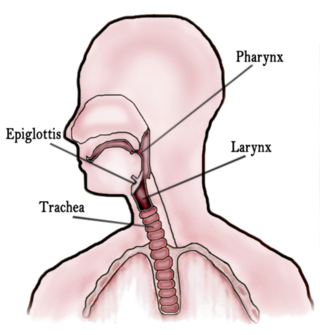
In vertebrate anatomy, the throat is the front part of the neck, internally positioned in front of the vertebrae. It contains the pharynx and larynx. An important section of it is the epiglottis, separating the esophagus from the trachea (windpipe), preventing food and drinks being inhaled into the lungs. The throat contains various blood vessels, pharyngeal muscles, the nasopharyngeal tonsil, the tonsils, the palatine uvula, the trachea, the esophagus, and the vocal cords. Mammal throats consist of two bones, the hyoid bone and the clavicle. The "throat" is sometimes thought to be synonymous for the fauces.
The laryngeal theory is a theory in historical linguistics positing that Proto-Indo-European had a number of laryngeal consonants that are not reconstructable by direct application of the comparative method to the Indo-European family. The 'missing' sounds remain consonants of an indeterminate place of articulation towards the back of the mouth, though further information is difficult to derive. Proponents aim to use the theory to:

In human anatomy, the subclavian arteries are paired major arteries of the upper thorax, below the clavicle. They receive blood from the aortic arch. The left subclavian artery supplies blood to the left arm and the right subclavian artery supplies blood to the right arm, with some branches supplying the head and thorax. On the left side of the body, the subclavian comes directly off the aortic arch, while on the right side it arises from the relatively short brachiocephalic artery when it bifurcates into the subclavian and the right common carotid artery.

The external carotid artery is a major artery of the head and neck. It arises from the common carotid artery when it splits into the external and internal carotid artery. The external carotid artery supplies blood to the face, brain and neck.

The recurrent laryngeal nerve (RLN) is a branch of the vagus nerve that supplies all the intrinsic muscles of the larynx, with the exception of the cricothyroid muscles. There are two recurrent laryngeal nerves, right and left. The right and left nerves are not symmetrical, with the left nerve looping under the aortic arch, and the right nerve looping under the right subclavian artery then traveling upwards. They both travel alongside the trachea. Additionally, the nerves are among the few nerves that follow a recurrent course, moving in the opposite direction to the nerve they branch from, a fact from which they gain their name.

The cricothyroid muscle is the only tensor muscle of the larynx aiding with phonation. It is innervated by the superior laryngeal nerve. Its action tilts the thyroid forward to help tense the vocal cords, thus increasing the pitch of the voice.
In anatomy, the left and right common carotid arteries (carotids) are arteries that supply the head and neck with oxygenated blood; they divide in the neck to form the external and internal carotid arteries.

The superior thoracic aperture, also known as the thoracic outlet, or thoracic inlet refers to the opening at the top of the thoracic cavity. It is also clinically referred to as the thoracic outlet, in the case of thoracic outlet syndrome. A lower thoracic opening is the inferior thoracic aperture.
Laryngeal consonants are consonants with their primary articulation in the larynx. The laryngeal consonants comprise the pharyngeal consonants, the glottal consonants, and for some languages uvular consonants.

The anterior jugular vein is a vein in the neck.

The inferior thyroid veins appear two, frequently three or four, in number, and arise in the venous plexus on the thyroid gland, communicating with the middle and superior thyroid veins. While the superior and middle thyroid veins serve as direct tributaries to the internal jugular vein, the inferior thyroid veins drain directly to the brachiocephalic veins.
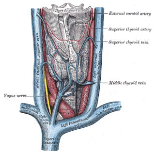
The superior thyroid vein is the vena comitans of the superior thyroid artery. It is formed by the union of deep and superficial tributaries that correspond to the arterial branches of the superior thyroid artery. Its tributaries are the superior laryngeal vein, and the cricothyroid veins. The vein empties into either the internal jugular vein, or the facial vein.

The carotid triangle is a portion of the anterior triangle of the neck.

The following outline is provided as an overview of and topical guide to human anatomy:
The superior laryngeal vein is a vein which drains the larynx into the superior thyroid vein.
Laryngeal vein can refer to:

The laryngeal tube is an airway management device designed as an alternative to other airway management techniques such as mask ventilation, laryngeal mask airway, and tracheal intubation. This device can be inserted blindly through the oropharynx into the hypopharynx to create an airway during anaesthesia and cardiopulmonary resuscitation so as to enable mechanical ventilation of the lungs.
