Related Research Articles

In anatomy, a sesamoid bone is a bone embedded within a tendon or a muscle. Its name is derived from the Greek word for 'sesame seed', indicating the small size of most sesamoids. Often, these bones form in response to strain, or can be present as a normal variant. The patella is the largest sesamoid bone in the body. Sesamoids act like pulleys, providing a smooth surface for tendons to slide over, increasing the tendon's ability to transmit muscular forces.

The levator labii superioris alaeque nasi muscle is, translated from Latin, the "lifter of both the upper lip and of the wing of the nose". The muscle is attached to the upper frontal process of the maxilla and inserts into the skin of the lateral part of the nostril and upper lip. At 44 characters, its name is longer than that of any other muscle.

The azygos vein is a vein running up the right side of the thoracic vertebral column draining itself towards the superior vena cava. It connects the systems of superior vena cava and inferior vena cava and can provide an alternative path for blood to the right atrium when either of the venae cavae is blocked.

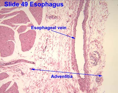
Esophageal varices are extremely dilated sub-mucosal veins in the lower third of the esophagus. They are most often a consequence of portal hypertension, commonly due to cirrhosis. People with esophageal varices have a strong tendency to develop severe bleeding which left untreated can be fatal. Esophageal varices are typically diagnosed through an esophagogastroduodenoscopy.

Portal hypertension is defined as increased portal venous pressure, with a hepatic venous pressure gradient greater than 5 mmHg. Normal portal pressure is 1–4 mmHg; clinically insignificant portal hypertension is present at portal pressures 5–9 mmHg; clinically significant portal hypertension is present at portal pressures greater than 10 mmHg. The portal vein and its branches supply most of the blood and nutrients from the intestine to the liver.

The mediastinum is the central compartment of the thoracic cavity. Surrounded by loose connective tissue, it is an undelineated region that contains a group of structures within the thorax, namely the heart and its vessels, the esophagus, the trachea, the phrenic and cardiac nerves, the thoracic duct, the thymus and the lymph nodes of the central chest.

In the human body, the femoral vein is the vein that accompanies the femoral artery in the femoral sheath. It is a deep vein that begins at the adductor hiatus as the continuation of the popliteal vein. The great saphenous vein, and the deep femoral vein drain into the femoral vein in the femoral triangle when it becomes known as the common femoral vein. It ends at the inferior margin of the inguinal ligament where it becomes the external iliac vein. Its major tributaries are the deep femoral vein, and the great saphenous vein. The femoral vein contains valves.

In anatomy, the fibular veins are accompanying veins of the fibular artery.

The superior thoracic aperture, also known as the thoracic outlet, or thoracic inlet refers to the opening at the top of the thoracic cavity. It is also clinically referred to as the thoracic outlet, in the case of thoracic outlet syndrome. A lower thoracic opening is the inferior thoracic aperture.

The hemiazygos vein is a vein running superiorly in the lower thoracic region, just to the left side of the vertebral column.

The anterior nasal spine, or anterior nasal spine of maxilla, is a bony projection in the skull that serves as a cephalometric landmark. The anterior nasal spine is the projection formed by the fusion of the two maxillary bones at the intermaxillary suture. It is placed at the level of the nostrils, at the uppermost part of the philtrum. It rarely fractures.

The appendicular artery, also known as the appendiceal artery, commonly arises from the terminal branch of the ileocolic artery, or less commonly from the posterior cecal artery or an ileal artery. It descends behind the termination of the ileum and enters the mesoappendix of the vermiform appendix. It runs near the free margin of the mesoappendix and ends in branches which supply the appendix.
In the course of the round ligament of the liver, small paraumbilical veins are found which establish an anastomosis between the veins of the anterior abdominal wall and the portal vein, hypogastric, and iliac veins. These veins include Burrow's veins, and the veins of Sappey – superior veins of Sappey and the inferior veins of Sappey.

The left gastric vein is a vein that derives from tributaries draining the lesser curvature of the stomach.
A portacaval anastomosis or portocaval anastomosis is a specific type of circulatory anastomosis that occurs between the veins of the portal circulation and the vena cava, thus forming one of the principal types of portasystemic anastomosis or portosystemic anastomosis, as it connects the portal circulation to the systemic circulation, providing an alternative pathway for the blood. When there is a blockage of the portal system, portocaval anastomosis enables the blood to still reach the systemic venous circulation. The inferior end of the esophagus and the superior part of the rectum are potential sites of a harmful portocaval anastomosis.

In human anatomy, an azygos lobe is a normal anatomical variation of the upper lobe of the right lung. It is seen in 0.3% of the population. Embryologically, it arises from an anomalous lateral course of the azygos vein, in a pleural septum within the apical segment of the right upper lobe or in other words an azygos lobe is formed when the right posterior cardinal vein, one of the precursors of the azygos vein, fails to migrate over the apex of the lung and penetrates it instead, carrying along two pleural layers as the azygous fissure, that invaginates into the upper portion of the right upper lobe.
Radiopaedia is a wiki-based international collaborative educational web resource containing a radiology encyclopedia and imaging case repository. It is currently the largest freely available radiology related resource in the world with more than 50,000 patient cases and over 16,000 reference articles on radiology-related topics. The open edit nature of articles allows radiologists, radiology trainees, radiographers, sonographers, and other healthcare professionals interested in medical imaging to refine most content through time. An editorial board peer reviews all contributions.

A developmental venous anomaly is a congenital variant of the cerebral venous drainage. On imaging it is seen as a number of small deep parenchymal veins converging toward a larger collecting vein.

A liver segment is one of eight segments of the liver as described in the widely used Couinaud classification in the anatomy of the liver. This system divides the lobes of the liver into eight segments based on a transverse plane through the bifurcation of the main portal vein, arranged in a clockwise manner starting from the caudate lobe.
The septal veins, also called the anterior septal veins and the veins of the septum pellucidum, are veins of the cerebral venous system which drain blood from the septum pellucidum of the anterior frontal lobe. The septal veins unify with the superior thalamostriate vein and the superior choridal vein at the interventricular foramina to form the internal cerebral veins.
References
![]() This article incorporates text in the public domain from the 20th edition of Gray's Anatomy (1918)
This article incorporates text in the public domain from the 20th edition of Gray's Anatomy (1918)
- ↑ Daniel J Bell; Henry Knipe; et al. "Azygos venous system | Radiology Reference Article | Radiopaedia.org". radiopaedia.org. Retrieved 2018-12-03.
- ↑ Daniel J Bell; Henry Knipe; et al. "Oesophagus | Radiology Reference Article | Radiopaedia.org". radiopaedia.org. Retrieved 2018-12-03.