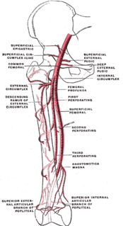
The femoral artery is a large artery in the thigh and the main arterial supply to the thigh and leg. It enters the thigh from behind the inguinal ligament as the continuation of the external iliac artery.

The femoral triangle is an anatomical region of the upper third of the thigh. It is a subfascial space which appears as a triangular depression below the inguinal ligament when the thigh is flexed, abducted and laterally rotated.

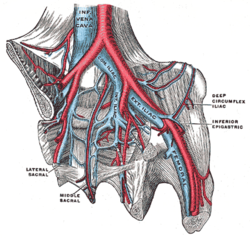
The external iliac arteries are two major arteries which bifurcate off the common iliac arteries anterior to the sacroiliac joint of the pelvis. They proceed anterior and inferior along the medial border of the psoas major muscles. They exit the pelvic girdle posterior and inferior to the inguinal ligament about one third laterally from the insertion point of the inguinal ligament on the pubic tubercle at which point they are referred to as the femoral arteries. The external iliac artery is usually the artery used to attach the renal artery to the recipient of a kidney transplant.
In human anatomy, iliac vein refers to several anatomical structures located in the pelvis:

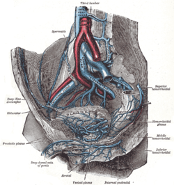
The internal iliac artery is the main artery of the pelvis.

The iliolumbar artery is the first branch of the posterior trunk of the internal iliac artery.

The superior gluteal artery is the largest branch of the internal iliac artery, and appears to be the continuation of the posterior division of that vessel. It is a short artery which runs backward between the lumbosacral trunk and the first sacral nerve, and divides into a superficial and a deep branch after passing out of the pelvis above the upper border of the piriformis muscle.
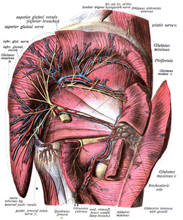
The inferior gluteal veins, or venæ comitantes of the inferior gluteal artery, begin on the upper part of the back of the thigh, where they anastomose with the medial femoral circumflex and first perforating veins.

The median sacral artery is a small vessel that arises posterior to the abdominal aorta and superior to its bifurcation.

The lateral circumflex femoral artery is an artery in the upper thigh.
The lumbar arteries are arteries located in the lower back or lumbar region. The lumbar arteries are in parallel with the intercostals.
The cruciate anastomosis is a circulatory anastomosis in the upper thigh of the inferior gluteal artery, the lateral and medial circumflex femoral arteries, and the first perforating artery of the profunda femoris artery. Also, the anastomotic branch of the posterior branch of the obturator artery. The cruciate anastomosis is clinically relevant because if there is a blockage between the femoral artery and external iliac artery, blood can reach the popliteal artery by means of the anastomosis. The route of blood is through the internal iliac, to the inferior gluteal artery, to a perforating branch of the deep femoral artery, to the lateral circumflex femoral artery, then to its descending branch into the superior lateral genicular artery and thus into the popliteal artery.

The internal iliac vein begins near the upper part of the greater sciatic foramen, passes upward behind and slightly medial to the internal iliac artery and, at the brim of the pelvis, joins with the external iliac vein to form the common iliac vein.

The deep circumflex iliac artery is an artery in the pelvis that travels along the iliac crest of the pelvic bone.
The superficial iliac circumflex artery, the smallest of the cutaneous branches of the femoral artery, arises close to the superficial epigastric artery, and, piercing the fascia lata, runs lateralward, parallel with the inguinal ligament, as far as the crest of the ilium.

The superficial external pudendal artery is one of the three pudendal arteries. It arises from the medial side of the femoral artery, close to the superficial epigastric artery and superficial iliac circumflex artery.
A circulatory anastomosis is a connection between two blood vessels, such as between arteries, between veins or between an artery and a vein. Anastomoses between arteries and between veins result in a multitude of arteries and veins, respectively, serving the same volume of tissue. Such anastomoses occur normally in the body in the circulatory system, serving as backup routes for blood to flow if one link is blocked or otherwise compromised, but may also occur pathologically.

The following outline is provided as an overview of and topical guide to human anatomy:
Iliac circumflex or Circumflex iliac can refer to:
The public domain consists of all the creative works to which no exclusive intellectual property rights apply. Those rights may have expired, been forfeited, expressly waived, or may be inapplicable.

Gray's Anatomy is an English language textbook of human anatomy originally written by Henry Gray and illustrated by Henry Vandyke Carter. Earlier editions were called Anatomy: Descriptive and Surgical, Anatomy of the Human Body and Gray's Anatomy: Descriptive and Applied, but the book's name is commonly shortened to, and later editions are titled, Gray's Anatomy. The book is widely regarded as an extremely influential work on the subject, and has continued to be revised and republished from its initial publication in 1858 to the present day. The latest edition of the book, the 41st, was published in September 2015.