Inferior vena cava

Last updated

Inferior vena cava
Diagram of the human heart (cropped).svg
Anterior (frontal) view of the opened heart. White arrows indicate valid blood flow.
Gray577.png
Superior vena cava, inferior vena cava, azygos vein and their tributaries
Details
Source common iliac vein
lumbar veins
testicular vein
renal vein
suprarenal vein
hepatic vein
Drains to Right atrium
Artery abdominal aorta
Identifiers
Latin vena cava inferior
Acronym(s)IVC
MeSH D014682
TA98 A12.3.09.001
TA2 4991
FMA 10951
Anatomical terminology

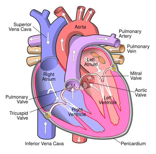
The inferior vena cava is a large vein that carries the deoxygenated blood from the lower and middle body into the right atrium of the heart. It is formed by the joining of the right and the left common iliac veins, usually at the level of the fifth lumbar vertebra. [1] [2]

Contents

The inferior vena cava is the lower ("inferior") of the two venae cavae, the two large veins that carry deoxygenated blood from the body to the right atrium of the heart: the inferior vena cava carries blood from the lower half of the body whilst the superior vena cava carries blood from the upper half of the body. Together, the venae cavae (in addition to the coronary sinus, which carries blood from the muscle of the heart itself) form the venous counterparts of the aorta.

It is a large retroperitoneal vein that lies posterior to the abdominal cavity and runs along the right side of the vertebral column. [1] It enters the right auricle at the lower right, back side of the heart. The name derives from Latin : vena, "vein", cavus, "hollow".

Structure

The IVC is formed by the joining of the left and right common iliac veins and brings collected blood into the right atrium of the heart. [1] It also joins with the azygos vein (which runs on the right side of the vertebral column) and venous plexuses next to the spinal cord.

The inferior vena cava begins as the left and right common iliac veins behind the abdomen unite, at about the level of L5. [1] [2] It passes through the thoracic diaphragm at the caval opening at the level of T8 - T9. [1] [3] It passes to the right of the descending aorta. [3]

Tributaries

The specific levels of the tributaries are as follows:

LevelVein
T8 hepatic veins, inferior phrenic vein
L1right suprarenal vein, renal veins
L2right gonadal vein
L1–L5 lumbar veins
L5 common iliac veins

Because the inferior vena cava is located to the right of the midline, drainage of the tributaries is not always symmetrical. On the right, the gonadal veins and suprarenal veins drain into the inferior vena cava directly. [1] On the left, they drain into the renal vein which in turn drains into the inferior vena cava. [1] By contrast, all the lumbar veins and hepatic veins usually drain directly into the inferior vena cava. [1]

Development

In the embryo, the inferior vena cava and right auricle are separated by the valve of the inferior vena cava, also known as the Eustachian valve. In the adult, this valve typically has totally regressed or remains as a small fold of endocardium. [4]

Variation

Rarely, the inferior vena cava may vary in its size and position. In transposition of the great arteries the inferior vena cava may lie on the left.

In between 0.2% to 0.3% of people, [5] the inferior vena cava may be duplicated beneath the level of the renal veins. [6]

Function

The inferior vena cava is a vein. It carries deoxygenated blood from the lower half of the body to the right atrium of the heart. [6]

The corresponding vein that carries deoxygenated blood from the upper half of the body is the superior vena cava.

Clinical significance

Health problems attributed to the IVC are most often associated with it being compressed (ruptures are rare because it has a low intraluminal pressure). Typical sources of external pressure are an enlarged aorta (abdominal aortic aneurysm), the gravid uterus (aortocaval compression syndrome) and abdominal malignancies, such as colorectal cancer, renal cell carcinoma and ovarian cancer. Since the inferior vena cava is primarily a right-sided structure, unconscious pregnant women should be turned on to their left side (the recovery position), to relieve pressure on it and facilitate venous return[ citation needed ]. In rare cases, straining associated with defecation can lead to restricted blood flow through the IVC and result in syncope (fainting). [7]

Blockage of the inferior vena cava is rare and is treated urgently as a life-threatening condition. It is associated with deep vein thrombosis, IVC filters, liver transplantation and surgical procedures such as the insertion of a catheter in the femoral vein in the groin. [8]

Branches of Inferior Vena Cava Major Veins.png
Branches of Inferior Vena Cava

Trauma to the vena cava is usually fatal as unstoppable excessive blood loss occurs.

Additional images

See also

Related Research Articles

<span class="mw-page-title-main">Vein</span> Blood vessels that carry blood towards the heart

Veins are blood vessels in the circulatory system of humans and most other animals that carry blood towards the heart. Most veins carry deoxygenated blood from the tissues back to the heart; exceptions are those of the pulmonary and fetal circulations which carry oxygenated blood to the heart. In the systemic circulation, arteries carry oxygenated blood away from the heart, and veins return deoxygenated blood to the heart, in the deep veins.

<span class="mw-page-title-main">Circulatory system</span> Organ system for circulating blood in animals

The circulatory system is a system of organs that includes the heart, blood vessels, and blood which is circulated throughout the entire body of a human or other vertebrate. It includes the cardiovascular system, or vascular system, that consists of the heart and blood vessels. The circulatory system has two divisions, a systemic circulation or circuit, and a pulmonary circulation or circuit. Some sources use the terms cardiovascular system and vascular system interchangeably with circulatory system.

<span class="mw-page-title-main">Superior vena cava</span> One of two veinous trunks bringing deoxygenated blood back to the heart

The superior vena cava (SVC) is the superior of the two venae cavae, the great venous trunks that return deoxygenated blood from the systemic circulation to the right atrium of the heart. It is a large-diameter (24 mm) short length vein that receives venous return from the upper half of the body, above the diaphragm. Venous return from the lower half, below the diaphragm, flows through the inferior vena cava. The SVC is located in the anterior right superior mediastinum. It is the typical site of central venous access via a central venous catheter or a peripherally inserted central catheter. Mentions of "the cava" without further specification usually refer to the SVC.

<span class="mw-page-title-main">Azygos vein</span> Human blood vessel by the spine

The azygos vein is a vein running up the right side of the thoracic vertebral column draining itself towards the superior vena cava. It connects the systems of superior vena cava and inferior vena cava and can provide an alternative path for blood to the right atrium when either of the venae cavae is blocked.

<span class="mw-page-title-main">Abdominal aorta</span> Largest artery in the abdomen

In human anatomy, the abdominal aorta is the largest artery in the abdominal cavity. As part of the aorta, it is a direct continuation of the descending aorta.

<span class="mw-page-title-main">Jugular vein</span> Veins that bring deoxygenated blood from the head back to the heart

The jugular veins are veins that take blood from the head back to the heart via the superior vena cava. The internal jugular vein descends next to the internal carotid artery and continues posteriorly to the sternocleidomastoid muscle.

<span class="mw-page-title-main">Atrium (heart)</span> Part of the human heart

The atrium is one of the two upper chambers in the heart that receives blood from the circulatory system. The blood in the atria is pumped into the heart ventricles through the atrioventricular mitral and tricuspid heart valves.

<span class="mw-page-title-main">Renal vein</span> Short thick veins which return blood from the kidneys to the vena cava

The renal veins in the renal circulation, are large-calibre veins that drain blood filtered by the kidneys into the inferior vena cava. There is one renal vein draining each kidney. Each renal vein is formed by the convergence of the interlobar veins of one kidney.

<span class="mw-page-title-main">Common iliac vein</span> Veins draining blood from the pelvis and lower limbs

In human anatomy, the common iliac veins are formed by the external iliac veins and internal iliac veins. The left and right common iliac veins come together in the abdomen at the level of the fifth lumbar vertebra, forming the inferior vena cava. They drain blood from the pelvis and lower limbs.

<span class="mw-page-title-main">Fetal circulation</span> Circulatory system of fetuses

In humans, the circulatory system is different before and after birth. The fetal circulation is composed of the placenta, umbilical blood vessels encapsulated by the umbilical cord, heart and systemic blood vessels. A major difference between the fetal circulation and postnatal circulation is that the lungs are not used during the fetal stage resulting in the presence of shunts to move oxygenated blood and nutrients from the placenta to the fetal tissue. At birth, the start of breathing and the severance of the umbilical cord prompt various changes that quickly transform fetal circulation into postnatal circulation.

<span class="mw-page-title-main">Coronary sinus</span> Set of veins which drain blood from the myocardium (heart muscle)

The coronary sinus is the largest vein of the heart. It drains over half of the deoxygenated blood from the heart muscle into the right atrium. It begins on the backside of the heart, in between the left atrium, and left ventricle; it begins at the junction of the great cardiac vein, and oblique vein of the left atrium. It receives multiple tributaries. It passes across the backside of the heart along a groove between left atrium and left ventricle, then drains into the right atrium at the orifice of the coronary sinus.

The term Great veins can refer to either —

<span class="mw-page-title-main">Hemiazygos vein</span>

The hemiazygos vein is a vein running superiorly in the lower thoracic region, just to the left side of the vertebral column.

<span class="mw-page-title-main">Hepatic portal system</span> System of veins comprising the hepatic portal vein and its tributaries

In human anatomy, the hepatic portal system or portal venous system is the system of veins comprising the portal vein and its tributaries. The other portal venous systems in the body are the renal portal system, and the hypophyseal portal system.

<span class="mw-page-title-main">Valve of inferior vena cava</span>

The valve of the inferior vena cava is a venous valve that lies at the junction of the inferior vena cava and right atrium.

<span class="mw-page-title-main">Ovarian vein</span>

The ovarian vein, the female gonadal vein, carries deoxygenated blood from its corresponding ovary to inferior vena cava or one of its tributaries. It is the female equivalent of the testicular vein, and is the venous counterpart of the ovarian artery. It can be found in the suspensory ligament of the ovary.

<span class="mw-page-title-main">Testicular vein</span>

The testicular vein, the male gonadal vein, carries deoxygenated blood from its corresponding testis to the inferior vena cava or one of its tributaries. It is the male equivalent of the ovarian vein, and is the venous counterpart of the testicular artery.

<span class="mw-page-title-main">Lumbar veins</span> Veins that drain the posterior abdominal wall

The lumbar veins are four pairs of veins running along the inside of the posterior abdominal wall, and drain venous blood from parts of the abdominal wall. Each lumbar vein accompanies a single lumbar artery. The lower two pairs of lumbar veins all drain directly into the inferior vena cava, whereas the fate of the upper two pairs is more variable.

<span class="mw-page-title-main">Venae cavae</span> Large veins which return blood from the body into the heart

In anatomy, the venae cavae are two large veins that return deoxygenated blood from the body into the heart. In humans they are the superior vena cava and the inferior vena cava, and both empty into the right atrium. They are located slightly off-center, toward the right side of the body.

The heart is a muscular organ situated in the mediastinum. It consists of four chambers, four valves, two main arteries, and the conduction system. The left and right sides of the heart have different functions: the right side receives de-oxygenated blood through the superior and inferior venae cavae and pumps blood to the lungs through the pulmonary artery, and the left side receives saturated blood from the lungs.

References

  1. 1 2 3 4 5 6 7 8 Mozes, GEZA; Gloviczki, PETER (January 1, 2007), Bergan, John J. (ed.), "CHAPTER 2 - Venous Embryology and Anatomy", The Vein Book, Burlington: Academic Press, pp. 15–25, doi:10.1016/b978-012369515-4/50005-3, ISBN   978-0-12-369515-4 , retrieved November 22, 2020
  2. 1 2 Dardis, Ronan M.; Saxena, Amar; Shad, Amjad; Chitnavis, Bhupal; Gullan, Richard (January 1, 2012), Quiñones-Hinojosa, Alfredo (ed.), "Chapter 154 - Disc Replacement Technologies in the Cervical and Lumbar Spine", Schmidek and Sweet Operative Neurosurgical Techniques (Sixth Edition), Philadelphia: W.B. Saunders, pp. 1777–1788, doi:10.1016/b978-1-4160-6839-6.10154-6, ISBN   978-1-4160-6839-6 , retrieved November 22, 2020
  3. 1 2 Blumgart, Leslie H.; Schwartz, Lawrence H.; DeMatteo, Ronald P. (January 1, 2017), Jarnagin, William R. (ed.), "Chapter 2 - Surgical and radiologic anatomy of the liver, biliary tract, and pancreas", Blumgart's Surgery of the Liver, Biliary Tract and Pancreas, 2-Volume Set (Sixth Edition), Philadelphia: Elsevier, pp. 32–59.e1, doi:10.1016/b978-0-323-34062-5.00002-9, ISBN   978-0-323-34062-5 , retrieved November 22, 2020
  4. Turhan Yavuz; Nazli, C; Kinay, O; Kutsal, A (2002). "Giant Eustachian Valve: with Echocardiographic Appearance of Divided Right Atrium". Texas Heart Institute Journal. 29 (4): 336–8. PMC   140300 . PMID   12484622.
  5. Stavropoulos, S. William; Solomon, Jeffrey A. (January 1, 2011), Pretorius, E. Scott; Solomon, Jeffrey A. (eds.), "Chapter 30 - Inferior Vena Cava Filters", Radiology Secrets Plus (Third Edition), Philadelphia: Mosby, pp. 223–227, doi:10.1016/b978-0-323-06794-2.00030-4, ISBN   978-0-323-06794-2 , retrieved November 22, 2020
  6. 1 2 Susan Standring; Neil R. Borley; et al., eds. (2008). Gray's anatomy : the anatomical basis of clinical practice (40th ed.). London: Churchill Livingstone. ISBN   978-0-8089-2371-8.
  7. Brophy, CM; Evans, L; Sumpio, BE (1993). "Defecation syncope secondary to functional inferior vena caval obstruction during a Valsalva maneuver". Annals of Vascular Surgery. 7 (4): 374–7. doi: 10.1007/BF02002893 . PMID   8268080. S2CID   42135883.
  8. Geehan DM, Inferior Vena Caval Thrombosis, emedicine.com, URL: http://www.emedicine.com/med/topic2718.htm, Accessed: August 3, 2005.