The femoral vein bears valves which are mostly bicuspid and whose number is variable between individuals and often between left and right leg.[1]
Course
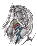
Veins of the leg. Common femoral vein shown, in common usage but not listed in TA.
The femoral vein continues into the thigh as the continuation from the popliteal vein at the back of the knee. It drains blood from the deep thigh muscles and thigh bone.[2] Proximal to the confluence with the deep femoral vein, and the joining of the great saphenous vein, the femoral vein is widely known as the common femoral vein.[3] As the common femoral vein leaves the inguinal ligament region it becomes the external iliac vein.[4] Other tributaries of the femoral vein are lateral and medial circumflex femoral veins.
The common femoral vein is the segment of the femoral vein between the branching point of the deep femoral vein and the inferior margin of the inguinal ligament.[5][6] It is not listed in Terminologia Anatomica, which is the international standard for human anatomical terminology developed by the Federative International Programme on Anatomical Terminology. However, it was thought to be due for inclusion in the next edition following consensus documents presented in 2001 at the 14th World Congress of the International Union of Phlebology, and in 2004 at the 21st World Congress of the International Union of Angiology.[7][8] These consensus documents were brought about by the need felt for more clarity and expansion of terms.[9][10]
Because of the widespread misunderstanding, and possible harmful results from the use of superficial femoral vein, a consensus was arrived at in 2001 during the World Congress of the International Union of Phlebology to change the name from superficial femoral vein simply to femoral vein.[13] This has been widely recognised and adopted though the use of superficial femoral vein still persists in some sources. Its use is actively discouraged.[14][15][16] It has been suggested that another term be used – the subsartorial vein.[17][18] A previous usage of subsartorial artery was published to avoid the name superficial femoral vein from being used.[19] As per the consensus of 2002, the superficial femoral artery was unchanged.[20]
The femoral vein is a common site for a deep vein thrombosis. This can be a proximal DVT in the femoral vein, or more proximal as an iliofemoral DVT usually associated with the common femoral vein. An iliofemoral DVT carries a greater risk of a pulmonary embolism developing.[22]
↑ Kachlik, D; Pechacek, V; Baca, V; Musil, V (June 2010). "The superficial venous system of the lower extremity: new nomenclature". Phlebology. 25 (3): 113–23. doi:10.1258/phleb.2009.009046. PMID20483860. S2CID8747016.
↑ Thiagarajah R, Venkatanarasimha N, Freeman S (2011). "Use of the term "superficial femoral vein" in ultrasound". J Clin Ultrasound. 39 (1): 32–34. doi:10.1002/jcu.20747. PMID20957733. S2CID23215861.
↑ Bundens WP, Bergan JJ, Halasz NA, Murray J, Drehobl M (October 1995). "The superficial femoral vein. A potentially lethal misnomer". JAMA. 274 (16): 1296–8. doi:10.1001/jama.1995.03530160048032. PMID7563535.
Anatomy figure: 12:05-01 at Human Anatomy Online, SUNY Downstate Medical Center—Veins of the lower extremity shown in association with major landmarks."
This page is based on this Wikipedia article Text is available under the CC BY-SA 4.0 license; additional terms may apply. Images, videos and audio are available under their respective licenses.