
The portal vein or hepatic portal vein is a blood vessel that carries blood from the gastrointestinal tract, gallbladder, pancreas and spleen to the liver. This blood contains nutrients and toxins extracted from digested contents. Approximately 75% of total liver blood flow is through the portal vein, with the remainder coming from the hepatic artery proper. The blood leaves the liver to the heart in the hepatic veins.

The pulmonary veins are the veins that transfer oxygenated blood from the lungs to the heart. The largest pulmonary veins are the four main pulmonary veins, two from each lung that drain into the left atrium of the heart. The pulmonary veins are part of the pulmonary circulation.

De humani corporis fabrica libri septem is a set of books on human anatomy written by Andreas Vesalius (1514–1564) and published in 1543. It was a major advance in the history of anatomy over the long-dominant work of Galen, and presented itself as such.

The celiacartery, also known as the celiac trunk, or truncus coeliacus, is the first major branch of the abdominal aorta. It is 1.25 cm in length. Branching from the aorta at thoracic vertebra 12 (T12) in humans, it is one of three anterior/ midline branches of the abdominal aorta.

In human anatomy, the superior mesenteric artery (SMA) arises from the anterior surface of the abdominal aorta, just inferior to the origin of the celiac trunk, and supplies the intestine from the lower part of the duodenum through two-thirds of the transverse colon, as well as the pancreas.
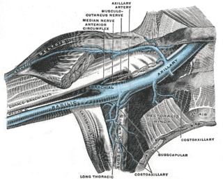
In human anatomy, the axillary vein is a large blood vessel that conveys blood from the lateral aspect of the thorax, axilla (armpit) and upper limb toward the heart. There is one axillary vein on each side of the body.
In human anatomy, the inferior mesenteric vein (IMV) is a blood vessel that drains blood from the large intestine. It usually terminates when reaching the splenic vein, which goes on to form the portal vein with the superior mesenteric vein (SMV). Anatomical variations include the IMV draining into the confluence of the SMV and splenic vein and the IMV draining in the SMV.

The facial vein is a relatively large vein in the human face. It commences at the side of the root of the nose and is a direct continuation of the angular vein where it also receives a small nasal branch. It lies behind the facial artery and follows a less tortuous course. It receives blood from the external palatine vein before it either joins the anterior branch of the retromandibular vein to form the common facial vein, or drains directly into the internal jugular vein.
The superficial temporal vein is a vein of the side of the head. It begins on the side and vertex of the skull in a network of veins which communicates with the frontal vein and supraorbital vein, with the corresponding vein of the opposite side, and with the posterior auricular vein and occipital vein. It ultimately crosses the posterior root of the zygomatic arch, enters the parotid gland, and unites with the internal maxillary vein to form the posterior facial vein.
The retromandibular vein, formed by the union of the superficial temporal and maxillary veins, descends in the substance of the parotid gland, superficial to the external carotid artery but beneath the facial nerve, between the ramus of the mandible and the sternocleidomastoideus muscle.

The internal pudendal veins are a set of veins in the pelvis. They are the venae comitantes of the internal pudendal artery.
The plantar digital veins arise from plexuses on the plantar surfaces of the digits, and, after sending intercapitular veins to join the dorsal digital veins, unite to form four metatarsal veins.

The vorticose veins, referred to clinically as the vortex veins, drain the ocular choroid. The number of vortex veins is known to vary from 4 to 8 with about 65% of the normal population having 4 or 5. In most cases, there is at least one vortex vein in each quadrant. Typically, the entrances to the vortex veins in the outer layer of the choroid can be observed funduscopically and provide an important clinical landmarks identifying the ocular equator. However, the veins run posteriorly in the sclera exiting the eye well posterior to the equator.

The intercostal arteries are a group of arteries that supply the area between the ribs ("costae"), called the intercostal space. The highest intercostal artery is an artery in the human body that usually gives rise to the first and second posterior intercostal arteries, which supply blood to their corresponding intercostal space. It usually arises from the costocervical trunk, which is a branch of the subclavian artery. Some anatomists may contend that there is no supreme intercostal artery, only a supreme intercostal vein.

In human anatomy, the dorsal veins of the penis comprise the superficial dorsal vein of the penis, the deep dorsal vein of the penis, the cavernosal veins of the penis, and the para-arterial veins of the penis. Although the documentation of the deep dorsal vein has been in place, the other veins are named by Dr. Geng Long Hsu; Collectively, they are coined as the "erection-related veins," owing to their physiological and clinical relevance.

The plantar metatarsal veins run backward in the metatarsal spaces, collect blood from digital veins and communicate, by means of perforating veins, with the veins on the dorsum of the foot, and unite to form the deep plantar venous arch which lies alongside the plantar arterial arch.
The volar digital veins on each finger are connected to the dorsal digital veins by oblique intercapitular veins. They drain into a venous plexus which is situated over the thenar and hypothenar eminences and across the front of the wrist.

The palmar digital veins on each finger are connected to the dorsal digital veins by oblique intercapitular veins.
The superficial palmar arch is accompanied by a pair of venae comitantes which constitute the superficial palmar venous arch. It receives the veins corresponding to the branches of the superficial arterial arch: the common palmar digital veins.

