External pudendal veins

Last updated
External pudendal veins
Gray549.png
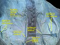
The left femoral triangle. (Superficial external pudendal vessels labeled at upper left.)
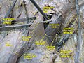
Details
Source Superficial dorsal vein of the penis/clitoris
Drains to Femoral vein
Artery External pudendal artery
Identifiers
Latin venae pudendae externae
TA98 A12.3.11.004
TA2 5059
FMA 70915
Anatomical terminology

The external pudendal veins (deep pudendal and superficial pudendal) are veins of the pelvis which drain into the great saphenous vein.

Additional images

See also


Related Research Articles

<span class="mw-page-title-main">Femoral artery</span> Large artery in the thigh

The femoral artery is a large artery in the thigh and the main arterial supply to the thigh and leg. The femoral artery gives off the deep femoral artery and descends along the anteromedial part of the thigh in the femoral triangle. It enters and passes through the adductor canal, and becomes the popliteal artery as it passes through the adductor hiatus in the adductor magnus near the junction of the middle and distal thirds of the thigh.

<span class="mw-page-title-main">Great saphenous vein</span> Large, subcutaneous, superficial vein of the leg

The great saphenous vein (GSV) or long saphenous vein is a large, subcutaneous, superficial vein of the leg. It is the longest vein in the body, running along the length of the lower limb, returning blood from the foot, leg and thigh to the deep femoral vein at the femoral triangle.

<span class="mw-page-title-main">Internal pudendal artery</span> Blood vessel supplying blood to the external genitalia

The internal pudendal artery is one of the three pudendal arteries. It branches off the internal iliac artery, and provides blood to the external genitalia.

<span class="mw-page-title-main">Superior mesenteric artery</span> Artery which supplies blood to the intestines and pancreas

In human anatomy, the superior mesenteric artery (SMA) is an artery which arises from the anterior surface of the abdominal aorta, just inferior to the origin of the celiac trunk, and supplies blood to the intestine from the lower part of the duodenum through two-thirds of the transverse colon, as well as the pancreas.

<span class="mw-page-title-main">Inferior mesenteric artery</span>

In human anatomy, the inferior mesenteric artery (IMA) is the third main branch of the abdominal aorta and arises at the level of L3, supplying the large intestine from the distal transverse colon to the upper part of the anal canal. The regions supplied by the IMA are the descending colon, the sigmoid colon, and part of the rectum.

<span class="mw-page-title-main">External iliac artery</span> Arteries of the pelvis

The external iliac arteries are two major arteries which bifurcate off the common iliac arteries anterior to the sacroiliac joint of the pelvis.

<span class="mw-page-title-main">Internal iliac artery</span> Main artery of the pelvis

The internal iliac artery is the main artery of the pelvis.

<span class="mw-page-title-main">Inferior epigastric artery</span> Blood vessel

In human anatomy, the inferior epigastric artery is an artery that arises from the external iliac artery. It is accompanied by the inferior epigastric vein; inferiorly, these two inferior epigastric vessels together travel within the lateral umbilical fold The inferior epigastric artery then traverses the arcuate line of rectus sheath to enter the rectus sheath, then anastomoses with the superior epigastric artery within the rectus sheath.

<span class="mw-page-title-main">Ilioinguinal nerve</span> Branch of the first lumbar nerve

The ilioinguinal nerve is a branch of the first lumbar nerve (L1). It separates from the first lumbar nerve along with the larger iliohypogastric nerve. It emerges from the lateral border of the psoas major just inferior to the iliohypogastric, and passes obliquely across the quadratus lumborum and iliacus. The ilioinguinal nerve then perforates the transversus abdominis near the anterior part of the iliac crest, and communicates with the iliohypogastric nerve between the transversus and the internal oblique muscle.

<span class="mw-page-title-main">Obturator nerve</span>

The obturator nerve in human anatomy arises from the ventral divisions of the second, third, and fourth lumbar nerves in the lumbar plexus; the branch from the third is the largest, while that from the second is often very small.

<span class="mw-page-title-main">Conjoint tendon</span> Medial part of the posterior wall of the inguinal canal

The conjoint tendon is a sheath of connective tissue formed from the lower part of the common aponeurosis of the abdominal internal oblique muscle and the transversus abdominis muscle, joining the muscle to the pelvis. It forms the medial part of the posterior wall of the inguinal canal.

<span class="mw-page-title-main">Pelvic cavity</span> Body cavity bounded by the pelvic bones

The pelvic cavity is a body cavity that is bounded by the bones of the pelvis. Its oblique roof is the pelvic inlet. Its lower boundary is the pelvic floor.

<span class="mw-page-title-main">Superficial epigastric artery</span> Large blood vessel

The superficial epigastric artery arises from the front of the femoral artery about 1 cm below the inguinal ligament, and, passing through the femoral sheath and the fascia cribrosa, turns upward in front of the inguinal ligament, and ascends between the two layers of the superficial fascia of the abdominal wall nearly as far as the umbilicus.

<span class="mw-page-title-main">Superficial circumflex iliac artery</span>

The superficial iliac circumflex artery, the smallest of the cutaneous branches of the femoral artery, arises close to the superficial epigastric artery, and, piercing the fascia lata, runs lateralward, parallel with the inguinal ligament, as far as the crest of the ilium.

<span class="mw-page-title-main">Superficial external pudendal artery</span>

The superficial external pudendal artery is one of the three pudendal arteries. It arises from the medial side of the femoral artery, close to the superficial epigastric artery and superficial iliac circumflex artery.

<span class="mw-page-title-main">Dorsal veins of the penis</span>

In human male anatomy, the dorsal veins of the penis are blood vessels that drain the shaft, the skin and the glans of the human penis. They are typically located in the midline on the dorsal aspect of the penis and they comprise the superficial dorsal veinof the penis, that lies in the subcutaneous tissue of the shaft, and the deep dorsal veinof the penis, that lies beneath the deep fascia.

<span class="mw-page-title-main">Superficial epigastric vein</span> Large blood vessel

The superficial epigastric vein is a vein which travels with the superficial epigastric artery. It joins the accessory saphenous vein near the fossa ovalis.

<span class="mw-page-title-main">Buck's fascia</span> Fascia in the penis

Buck's fascia is a layer of deep fascia covering the three erectile bodies of the penis.

<span class="mw-page-title-main">Saphenous opening</span>

In anatomy, the saphenous opening is an oval opening in the upper mid part of the fascia lata of the thigh. It lies 3–4 cm below and lateral to the pubic tubercle and is about 3 cm long and 1.5 cm wide.

<span class="mw-page-title-main">Anterior accessory saphenous vein</span> Large blood vessel

The anterior accessory saphenous vein is a special anterior tributary of the great saphenous vein (GSV), draining the antero-lateral face of the thigh.