Thigh

Last updated
Thigh
Thigh.jpg
A woman's thighs
Braus 1921 277.png
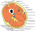
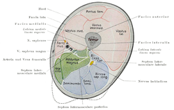
Cross-section of the thigh showing muscles and bone (latin terminology)
Details
Identifiers
Latin femur
MeSH D013848
TA98 A01.1.00.035
TA2 160
FMA 24967
Anatomical terminology

In anatomy, the thigh is the area between the hip (pelvis) and the knee. Anatomically, it is part of the lower limb. [1]

Contents

The single bone in the thigh is called the femur. This bone is very thick and strong (due to the high proportion of bone tissue), and forms a ball and socket joint at the hip, and a modified hinge joint at the knee. [2]

Structure

Bones

The femur is the only bone in the thigh and serves as an attachment site for all muscles in the thigh. The head of the femur articulates with the acetabulum in the pelvic bone forming the hip joint, while the distal part of the femur articulates with the tibia and patella forming the knee. By most measures, the femur is the strongest bone in the body. The femur is also the longest bone in the body. [3]

The femur is categorised as a long bone and comprises a diaphysis, the shaft (or body) and two epiphyses, the lower extremity and the upper extremity of femur, that articulate with adjacent bones in the hip and knee. [4]

Muscular compartments

In cross-section, the thigh is divided up into three separate compartments, divided by fascia, each containing muscles. These compartments use the femur as an axis and are separated by tough connective tissue membranes (or septa). Each of these compartments has its own blood and nerve supply, and contains a different group of muscles.

Anterior compartment muscles of the thigh include sartorius, and the four muscles that comprise the quadriceps musclesrectus femoris, vastus medialis, vastus intermedius and vastus lateralis.

Posterior compartment muscles of the thigh are the hamstring muscles, which include semimembranosus, semitendinosus, and biceps femoris.

Medial compartment muscles are pectineus, adductor magnus, adductor longus and adductor brevis, and also gracilis.

Because the major muscles of the thigh are the largest muscles of the body, resistance exercises (strength training) of them stimulate blood flow more than any other localized activity. [5]

Blood supply

Main blood vessels of the thigh. Common femoral and subsartorial artery and vein.jpg
Main blood vessels of the thigh.

The arterial supply is by the femoral artery and the obturator artery. The lymphatic drainage closely follows the arterial supply and drains to the lumbar lymphatic trunks on the corresponding side, which in turn drains to the cisterna chyli.

The deep venous system of the thigh consists of the femoral vein, common femoral vein, deep femoral vein, the proximal part of the popliteal vein, and various smaller vessels; these are the site of proximal deep vein thrombosis. The perforating veins connect the deep and the superficial system, which consists of the small and great saphenous veins (the site of varicose veins). [7]

Clinical significance

Thigh weakness can result in a positive Gowers' sign on physical examination. [8]

Thigh injury resulting from sports, whether acute or from overuse, can mean significant incapacity to perform. Soft tissue injury can encompass sprains, strains, bruising and tendinitis.

Runner's knee (patellofemoral pain) is a direct consequence of the kneecap rubbing against the end of the thigh bone (femur). Tight hamstrings and weak thigh muscles, required to stabilize the knee, increase the risk of developing of runner's knee. [9]

Society and culture

Western societies generally tolerate clothing that displays thighs, such as short shorts and miniskirts. Beachwear and many athleisure styles often display thighs as well. Professional dress codes may require covering up bare thighs.

Many Islamic countries disapprove of or prohibit the display of thighs, especially by women.

Strategic covering or display of thighs is used in popular fashion around the world, such as thigh-high boots and zettai ryoiki .

Additional images

Related Research Articles

<span class="mw-page-title-main">Human leg</span> Lower extremity or limb of the human body (foot, lower leg, thigh and hip)

The leg is the entire lower limb of the human body, including the foot, thigh or sometimes even the hip or buttock region. The major bones of the leg are the femur, tibia, and adjacent fibula. The thigh is between the hip and knee, while the calf (rear) and shin (front) are between the knee and foot.

<span class="mw-page-title-main">Femur</span> Thigh bone

The femur, or thigh bone is the only bone in the thigh. The thigh is the region of the lower limb between the hip and the knee. In many four-legged animals the femur is the upper bone of the hindleg.

<span class="mw-page-title-main">Deep femoral artery</span> Large branch of the femoral artery that supplies the thigh

The deep femoral artery also known as the deep artery of the thigh, or profunda femoris artery, is a large branch of the femoral artery. It travels more deeply ("profoundly") than the rest of the femoral artery. It gives rise to the lateral circumflex femoral artery and medial circumflex femoral artery, and the perforating arteries, terminating within the thigh.

<span class="mw-page-title-main">Pectineus muscle</span> Adductor of the thigh

The pectineus muscle is a flat, quadrangular muscle, situated at the anterior (front) part of the upper and medial (inner) aspect of the thigh. The pectineus muscle is the most anterior adductor of the hip. The muscle's primary action is hip flexion; it also produces adduction and internal rotation of the hip.

<span class="mw-page-title-main">Hip</span> Anatomical region between the torso and the legs, holding the buttocks and genital region

In vertebrate anatomy, the hip, or coxa(pl.: coxae) in medical terminology, refers to either an anatomical region or a joint on the outer (lateral) side of the pelvis.

The biceps femoris is a muscle of the thigh located to the posterior, or back. As its name implies, it consists of two heads; the long head is considered part of the hamstring muscle group, while the short head is sometimes excluded from this characterization, as it only causes knee flexion and is activated by a separate nerve.

<span class="mw-page-title-main">Adductor brevis muscle</span> Muscle in the thigh situated immediately behind the pectineus and adductor longus

The adductor brevis is a muscle in the thigh situated immediately deep to the pectineus and adductor longus. It belongs to the adductor muscle group. The main function of the adductor brevis is to pull the thigh medially. The adductor brevis and the rest of the adductor muscle group is also used to stabilize left to right movements of the trunk, when standing on both feet, or to balance when standing on a moving surface. The adductor muscle group is used pressing the thighs together to ride a horse, and kicking with the inside of the foot in soccer or swimming. Last, they contribute to flexion of the thigh when running or against resistance.

<span class="mw-page-title-main">Adductor longus muscle</span> Skeletal muscle located in the thigh

In the human body, the adductor longus is a skeletal muscle located in the thigh. One of the adductor muscles of the hip, its main function is to adduct the thigh and it is innervated by the obturator nerve. It forms the medial wall of the femoral triangle.

<span class="mw-page-title-main">Adductor magnus muscle</span> Muscle in the thigh

The adductor magnus is a large triangular muscle, situated on the medial side of the thigh.

<span class="mw-page-title-main">Gracilis muscle</span> Most superficial muscle on the medial side of the thigh

The gracilis muscle is the most superficial muscle on the medial side of the thigh. It is thin and flattened, broad above, narrow and tapering below.

The semimembranosus muscle is the most medial of the three hamstring muscles in the thigh. It is so named because it has a flat tendon of origin. It lies posteromedially in the thigh, deep to the semitendinosus muscle. It extends the hip joint and flexes the knee joint.

The rectus femoris muscle is one of the four quadriceps muscles of the human body. The others are the vastus medialis, the vastus intermedius, and the vastus lateralis. All four parts of the quadriceps muscle attach to the patella by the quadriceps tendon.

<span class="mw-page-title-main">Femoral nerve</span> Long nerve down the thigh and inner leg

The femoral nerve is a nerve in the thigh that supplies skin on the upper thigh and inner leg, and the muscles that extend the knee. It is the largest branch of the lumbar plexus.

<span class="mw-page-title-main">Muscles of the hip</span> Causes movement in the hip

In human anatomy, the muscles of the hip joint are those muscles that cause movement in the hip. Most modern anatomists define 17 of these muscles, although some additional muscles may sometimes be considered. These are often divided into four groups according to their orientation around the hip joint: the gluteal group; the lateral rotator group; the adductor group; and the iliopsoas group.

<span class="mw-page-title-main">Fascia lata</span> Deep fascia of the thigh

The fascia lata is the deep fascia of the thigh. It encloses the thigh muscles and forms the outer limit of the fascial compartments of thigh, which are internally separated by the medial intermuscular septum and the lateral intermuscular septum. The fascia lata is thickened at its lateral side where it forms the iliotibial tract, a structure that runs to the tibia and serves as a site of muscle attachment.

<span class="mw-page-title-main">Lateral circumflex femoral artery</span>

The lateral circumflex femoral artery is an artery in the upper thigh. It is usually a branch of the profunda femoris artery, and produces three branches. It is mostly distributed to the muscles of the lateral thigh, supplying arterial blood to muscles of the knee extensor group.

<span class="mw-page-title-main">Medial circumflex femoral artery</span>

The medial circumflex femoral artery is an artery in the upper thigh that arises from the profunda femoris artery. It supplies arterial blood to several muscles in the region, as well as the femoral head and neck.

<span class="mw-page-title-main">Posterior compartment of thigh</span> One of the fascial compartments that contains the knee flexors and hip extensors

The posterior compartment of the thigh is one of the fascial compartments that contains the knee flexors and hip extensors known as the hamstring muscles, as well as vascular and nervous elements, particularly the sciatic nerve.

<span class="mw-page-title-main">Anterior compartment of thigh</span> Muscles which extend the knee and flex the hip

The anterior compartment of thigh contains muscles which extend the knee and flex the hip.

<span class="mw-page-title-main">Outline of human anatomy</span> Overview of and topical guide to human anatomy

The following outline is provided as an overview of and topical guide to human anatomy:

References

  1. "thigh" at Dorland's Medical Dictionary [ dead link ]
  2. "Thigh". Cambridge Dictionary. Retrieved 2021-06-23.
  3. "Facts About Bones in Your Skeleton". Hospital for Special Surgery.
  4. Bojsen-Møller, Finn; Simonsen, Erik B.; Tranum-Jensen, Jørgen (2001). Bevægeapparatets anatomi[Anatomy of the Locomotive Apparatus] (in Danish) (12th ed.). pp. 239–241. ISBN   978-87-628-0307-7.
  5. Cespedes, Andrea. "How Important Are Leg Workouts for Muscle Gain?".
  6. Amarnath C and Hemant Patel (2023). Comprehensive Textbook of Clinical Radiology – Volume III: Chest and Cardiovascular system. Elsevier Health Sciences. p.  1072. ISBN   9788131263617.
  7. "Thigh". The Lecturio Medical Concept Library. Retrieved 2021-06-23.
  8. Chang, Richard F., MD; Mubarak, Scott J., MD (2011-12-28). "Pathomechanics of Gowers' Sign: A Video Analysis of a Spectrum of Gowers' Maneuvers". Clinical Orthopaedics and Related Research. 470 (7): 1987–1991. doi:10.1007/s11999-011-2210-6. PMC   3369091 . PMID   22203329.{{cite journal}}: CS1 maint: multiple names: authors list (link)
  9. "Medically Sound: Field, Court, and Course Related Injuries – Sports Injuries". Medically Sound. 2020-08-31. Retrieved 2020-11-01.