Gemelli muscles

Last updated
Gemelli muscles
Posterior Hip Muscles 1.PNG
The gemelli muscles and the internal obturator muscle
Details
Origin Ischiopubic ramus and obturator membrane
Anatomical terms of muscle

The gemelli muscles are the inferior gemellus muscle and the superior gemellus muscle, two small accessory fasciculi to the tendon of the internal obturator muscle. The gemelli muscles belong to the lateral rotator group of six muscles of the hip that rotate the femur in the hip joint.

Contents

Superior gemellus muscle

Gemelli muscles - superior shown in red above internal obturator, and inferior shown below the internal obturator. Gemellus superior muscle.PNG
Gemelli muscles – superior shown in red above internal obturator, and inferior shown below the internal obturator.

The gemelli muscles are two small muscular fasciculi, accessories to the tendon of the internal obturator muscle which is received into a groove between them. The superior gemellus muscle is the higher placed gemellus muscle that arises from the outer (gluteal) surface of the ischial spine, and blends with the upper part of the tendon of the internal obturator. It is smaller than the inferior gemellus. In some people, the fibres of the gemellus superior extend further than average, and are prolonged onto the medial surface of the greater trochanter of the femur. [1]

The superior and inferior gemelli are supplied by the inferior gluteal artery. Nerve supply to the superior gemellus is from the supply to the internal obturator – L5, S1, and S2. [2]

Inferior gemellus muscle

The inferior gemellus muscle arises from the upper part of the ischial tuberosity, immediately below the groove for the internal obturator tendon. It blends with the lower part of the tendon, and is inserted with it into the medial surface of the greater trochanter. It is rarely absent.

Like the internal obturator muscle, the gemellus superior and gemellus inferior help to steady the femoral head in the acetabulum. Both muscles also help to laterally rotate the extended thigh and abduct the flexed thigh at the hip by assisting the internal obturator. [1] The gemelli muscles act to compensate the reduced power of the internal obturator as it turns around the lesser sciatic notch. [1]

Blood supply is from the inferior gluteal artery. Nerve supply is from the supply to the quadratus femoris – L4 to S1. [2]

Etymology

Gemellus is the diminutive of "geminus"[ clarification needed ] meaning twin, doubled or duplicated. The superior and inferior gemellus muscles are paired and perform the same action.

Additional images

Related Research Articles

<span class="mw-page-title-main">Human leg</span> Lower extremity or limb of the human body (foot, lower leg, thigh and hip)

The leg is the entire lower limb of the human body, including the foot, thigh or sometimes even the hip or buttock region. The major bones of the leg are the femur, tibia, and adjacent fibula. The thigh is between the hip and knee, while the calf (rear) and shin (front) are between the knee and foot.

<span class="mw-page-title-main">Gluteus minimus</span> Smallest of the three gluteal muscles

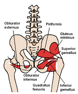
The gluteus minimus, or glutæus minimus, the smallest of the three gluteal muscles, is situated immediately beneath the gluteus medius.

<span class="mw-page-title-main">Greater trochanter</span> Quadrilateral part of the thigh bone

The greater trochanter of the femur is a large, irregular, quadrilateral eminence and a part of the skeletal system.

<span class="mw-page-title-main">Piriformis muscle</span> One of six small hip muscles in the lateral rotator group

The piriformis muscle is a flat, pyramidally-shaped muscle in the gluteal region of the lower limbs. It is one of the six muscles in the lateral rotator group.

<span class="mw-page-title-main">Pectineus muscle</span> Adductor of the thigh

The pectineus muscle is a flat, quadrangular muscle, situated at the anterior (front) part of the upper and medial (inner) aspect of the thigh. The pectineus muscle is the most anterior adductor of the hip. The muscle's primary action is hip flexion; it also produces adduction and internal rotation of the hip.

<span class="mw-page-title-main">Obturator internus muscle</span> One of six small hip muscles in the lateral rotator group

The internal obturator muscle or obturator internus muscle originates on the medial surface of the obturator membrane, the ischium near the membrane, and the rim of the pubis.

<span class="mw-page-title-main">Hip</span> Anatomical region between the torso and the legs, holding the buttocks and genital region

In vertebrate anatomy, the hip, or coxa(pl.: coxae) in medical terminology, refers to either an anatomical region or a joint on the outer (lateral) side of the pelvis.

The biceps femoris is a muscle of the thigh located to the posterior, or back. As its name implies, it consists of two heads; the long head is considered part of the hamstring muscle group, while the short head is sometimes excluded from this characterization, as it only causes knee flexion and is activated by a separate nerve.

<span class="mw-page-title-main">External obturator muscle</span> One of six small hip muscles in the lateral rotator group

The external obturator muscle, obturator externus muscle is a flat, triangular muscle, which covers the outer surface of the anterior wall of the pelvis.

<span class="mw-page-title-main">Adductor magnus muscle</span> Muscle in the thigh

The adductor magnus is a large triangular muscle, situated on the medial side of the thigh.

<span class="mw-page-title-main">Muscles of the hip</span> Causes movement in the hip

In human anatomy, the muscles of the hip joint are those muscles that cause movement in the hip. Most modern anatomists define 17 of these muscles, although some additional muscles may sometimes be considered. These are often divided into four groups according to their orientation around the hip joint: the gluteal group; the lateral rotator group; the adductor group; and the iliopsoas group.

<span class="mw-page-title-main">Inferior gluteal nerve</span>

The inferior gluteal nerve is the main motor neuron that innervates the gluteus maximus muscle. It is responsible for the movement of the gluteus maximus in activities requiring the hip to extend the thigh, such as climbing stairs. Injury to this nerve is rare but often occurs as a complication of posterior approach to the hip during hip replacement. When damaged, one would develop gluteus maximus lurch, which is a gait abnormality which causes the individual to 'lurch' backwards to compensate lack in hip extension.

<span class="mw-page-title-main">Ischium</span> Lower and back region of the hip bone

The ischium forms the lower and back region of the hip bone.

<span class="mw-page-title-main">Superior gluteal artery</span>

The superior gluteal artery is the terminal branch of the posterior division of the internal iliac artery. It exits the pelvis through the greater sciatic foramen before splitting into a superficial branch and a deep branch.

<span class="mw-page-title-main">Inferior gluteal artery</span>

The inferior gluteal artery is a terminal branch of the anterior trunk of the internal iliac artery. It exits the pelvis through the greater sciatic foramen. It is distributed chiefly to the buttock and the back of the thigh.

<span class="mw-page-title-main">Obturator artery</span>

The obturator artery is a branch of the internal iliac artery that passes antero-inferiorly on the lateral wall of the pelvis, to the upper part of the obturator foramen, and, escaping from the pelvic cavity through the obturator canal, it divides into an anterior branch and a posterior branch.

<span class="mw-page-title-main">Upper extremity of femur</span>

The upper extremity, proximal extremity or superior epiphysis of the femur is the part of the femur closest to the pelvic bone and the trunk. It contains the following structures:

<span class="mw-page-title-main">Trochanteric fossa</span>

In mammals including humans, the medial surface of the greater trochanter has at its base a deep depression bounded posteriorly by the intertrochanteric crest, called the trochanteric fossa. This fossa is the point of insertion of four muscles. Moving from the inferior-most to the superior-most, they are: the tendon of the obturator externus muscle, the obturator internus, the superior gemellus and inferior gemellus. The width and depth of the trochanteric fossa varies taxonomically.

<span class="mw-page-title-main">Outline of human anatomy</span> Overview of and topical guide to human anatomy

The following outline is provided as an overview of and topical guide to human anatomy:

<span class="mw-page-title-main">Hip bone</span> Bone of the pelvis

The hip bone is a large flat bone, constricted in the center and expanded above and below. In some vertebrates it is composed of three parts: the ilium, ischium, and the pubis.

References

  1. 1 2 3 Palastanga, Nigel; Soames, Roger (November 2011). Physiotherapy Essentials : Anatomy and Human Movement : Structure and Function (6th ed.). London, GBR: Elsevier Health Sciences. pp. 235–237. ISBN   9780702035531.
  2. 1 2 Miniato, Mohammed A.; Black, Asa C.; Varacallo, Matthew (2023), "Anatomy, Back, Lumbosacral Trunk", StatPearls, Treasure Island (FL): StatPearls Publishing, PMID   30969700 , retrieved 2023-12-28