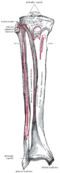
 Bones of the right leg, anterior surface
 Left calcaneus, inferior surface
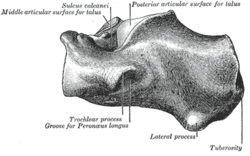 Left calcaneus, lateral surface
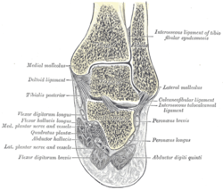 Coronal section through right talocrural and talocalcaneal joints
 Fibularis (peroneus) longus labeled at right
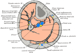 Cross-section through middle of leg
 The popliteal, posterior tibial, and fibular arteries
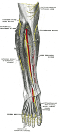 Deep nerves of the front of the leg
 Back of left lower extremity
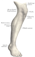 Lateral aspect of right leg
 Muscles of leg (lateral view, deep dissection)
| fibularis longus | |
|---|---|
 Animation | |
 Mucous sheaths of tendons around right ankle, lateral aspect. (Tendon-sheath of fibularis longus labeled as peronaeus longus at bottom center.) | |
| Details | |
| Origin | Proximal part of lateral surface of shaft of fibula [1] and head of fibula |
| Insertion | First metatarsal, medial cuneiform [1] |
| Artery | Fibular (peroneal) artery |
| Nerve | Superficial fibular nerve [1] |
| Actions | Plantarflexion, eversion, support arches [1] |
| Antagonist | Tibialis anterior muscle |
| Identifiers | |
| Latin | musculus fibularis longus |
| TA98 | A04.7.02.041 |
| TA2 | 2652 |
| FMA | 22539 |
| Anatomical terms of muscle | |
In human anatomy, the fibularis longus (also known as peroneus longus) is a superficial muscle in the lateral compartment of the leg. It acts to tilt the sole of the foot away from the midline of the body (eversion) and to extend the foot downward away from the body (plantar flexion) at the ankle.
Contents
The fibularis longus is the longest and most superficial of the three fibularis (peroneus) muscles. At its upper end, it is attached to the head of the fibula, and its "belly" runs down along most of this bone. The muscle becomes a tendon that wraps around and behind the lateral malleolus of the ankle, then continues under the foot to attach to the medial cuneiform and first metatarsal. It is supplied by the superficial fibular nerve.


