The fascia lata is an investment for the whole of the thigh, but varies in thickness in different parts. It is thicker in the upper and lateral part of the thigh, where it receives a fibrous expansion from the gluteus maximus, and where the tensor fasciae latae is inserted between its layers; it is very thin behind and at the upper and medial part, where it covers the adductor muscles, and again becomes stronger around the knee, receiving fibrous expansions from the tendon of the biceps femoris laterally, from the sartorius medially, and from the quadriceps femoris in front.
Function
The fascia lata surrounds the tensor fasciae latae muscle. It is a fibrous sheath that encircles the thigh subcutaneously. This encircling of the muscle allows the muscles to be bound together tightly.[citation needed]
From its attachment to the iliac crest it passes down over the gluteus medius to the upper border of the gluteus maximus, where it splits into two layers, one passing superficial to and the other beneath this muscle; at the lower border of the muscle the two layers reunite.
Laterally
Laterally, the fascia lata receives the greater part of the tendon of insertion of the gluteus maximus, and becomes proportionately thickened.
The portion of the fascia lata attached to the front part of the iliac crest, and corresponding to the origin of the tensor fasciae latae, extends down the lateral side of the thigh as two layers, one superficial to and the other beneath this muscle; at the lower end of the muscle these two layers unite and form a strong band, having first received the insertion of the muscle.
This band is continued downward under the name of the iliotibial band and is attached to the lateral condyle of the tibia.
The part of the iliotibial band which lies beneath the tensor fasciae latae is prolonged upward to join the lateral part of the capsule of the hip joint.
Below
Below, the fascia lata is attached to all the prominent points around the knee joint, viz., the condyles of the femur and tibia, and the head of the fibula.
On either side of the kneecap it is strengthened by transverse fibers from the lower parts of the vasti muscles (three of the four quadriceps) which are attached to and support this bone.
Of these the lateral are the stronger, and are continuous with the iliotibial band.
Since the 1920s fasciae latae from deceased donors have been used in reconstructive surgery. In 1999 preserved mashed fasciae latae became FDA-approved as a tissue product designed to replace areas of lost fascia or collagen.[3] The fascia lata normally performs the function of encircling and tightening the muscles in the thigh. Because of this function, it has been used as grafts for patients with facial paralysis. The fascia lata offers supports to the muscles that make up the face and this support increases the recovery of the facial muscles. The surgeons use the fascia lata as a sort of facial sling to support up the paralyzed face and loops the fascia lata around the center of the lower lip, the corner of the mouth and the center of the upper lip.[4] A small portion of fascia lata harvested through a sub centimeter skin incision on the lower lateral side of the thigh is used for reconstructing the ear drum in tympanoplasty surgery. A larger portion is used in nasal endoscopic skull base surgery.
History
Etymology
It is named from its great extent. "Latus" give the superlative "Latissimus" meaning broadest or widest.[5]
Additional images
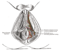
The superficial branches of the internal pudendal artery.
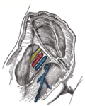
Femoral sheath laid open to show its three compartments.
This page is based on this Wikipedia article Text is available under the CC BY-SA 4.0 license; additional terms may apply. Images, videos and audio are available under their respective licenses.