Lateral rotator group

Last updated
Lateral rotator group
Posterior Hip Muscles 1.PNG
The lateral rotator group and the gluteus minimus muscle, posterior view
Gray344.png
Structures surrounding left hip joint
Details
Origin At or below the acetabulum of the ilium
Insertion On or near the greater trochanter of the femur
Artery Inferior gluteal artery, lateral sacral artery, superior gluteal artery
Nerve Obturator nerve, nerve to the piriformis, nerve to quadratus femoris
Actions Lateral rotation of hip
Antagonist Gluteus minimus muscle, gluteus medius muscle
Anatomical terms of muscle

The lateral rotator group is a group of six small muscles of the hip which all externally (laterally) rotate the femur in the hip joint. It consists of the following muscles: piriformis, gemellus superior, obturator internus, gemellus inferior, quadratus femoris and the obturator externus. [1]

Contents

All muscles in the lateral rotator group originate from the hip bone and insert on to the upper extremity of the femur. The muscles are innervated by the sacral plexus (L4-S2), except the obturator externus muscle, which is innervated by the lumbar plexus. [2]

Individual muscles

Muscleorigininsertioninnervation [2]
Piriformis muscle Anterior surface of sacrum between and laterally to the anterior sacral foramina Superior boundary of greater trochanter Nerve to the piriformis (S1-S2)
Gemellus superior muscle Ischial spine Upper edge of Obturator internus muscle tendon (indirectly greater trochanter) Nerve to obturator internus (L5-S2)
Obturator internus muscle Medial surface of obturator membrane and the surrounding bone Medial surface of greater trochanter Nerve to obturator internus (L5-S2)
Gemellus inferior muscle Just above the tuberosity of the ischium Lower edge of Obturator internus muscle tendon (indirectly greater trochanter) Nerve to quadratus femoris (L4-S1)
Quadratus femoris muscle Lateral edge of the tuberosity of the ischium Intertrochanteric crest Nerve to quadratus femoris (L4-S1)
Obturator externus muscle Lateral surface of obturator membrane and the ischiopubic ramus Trochanteric fossa Posterior branch of obturator nerve (L3-L4)

Other lateral rotators

This group does not include all muscles which aid in lateral rotation of the hip joint: rather it is a collection of ones which are known for primarily performing this action. Other muscles that contribute to lateral rotation of the hip include:

Additional images

See also

Related Research Articles

<span class="mw-page-title-main">Human leg</span> Lower extremity or limb of the human body (foot, lower leg, thigh and hip)

The leg is the entire lower limb of the human body, including the foot, thigh or sometimes even the hip or buttock region. The major bones of the leg are the femur, tibia, and adjacent fibula. The thigh is between the hip and knee, while the calf (rear) and shin (front) are between the knee and foot.

<span class="mw-page-title-main">Greater trochanter</span> Quadrilateral part of the thigh bone

The greater trochanter of the femur is a large, irregular, quadrilateral eminence and a part of the skeletal system.

<span class="mw-page-title-main">Piriformis muscle</span> One of six small hip muscles in the lateral rotator group

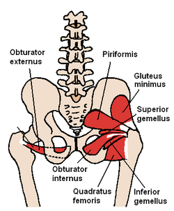
The piriformis muscle is a flat, pyramidally-shaped muscle in the gluteal region of the lower limbs. It is one of the six muscles in the lateral rotator group.

<span class="mw-page-title-main">Pectineus muscle</span> Adductor of the thigh

The pectineus muscle is a flat, quadrangular muscle, situated at the anterior (front) part of the upper and medial (inner) aspect of the thigh. The pectineus muscle is the most anterior adductor of the hip. The muscle's primary action is hip flexion; it also produces adduction and internal rotation of the hip.

<span class="mw-page-title-main">Hip</span> Anatomical region between the torso and the legs, holding the buttocks and genital region

In vertebrate anatomy, the hip, or coxa(pl.: coxae) in medical terminology, refers to either an anatomical region or a joint on the outer (lateral) side of the pelvis.

<span class="mw-page-title-main">Quadratus femoris muscle</span> One of six small hip muscles in the lateral rotator group

The quadratus femoris is a flat, quadrilateral skeletal muscle. Located on the posterior side of the hip joint, it is a strong external rotator and adductor of the thigh, but also acts to stabilize the femoral head in the acetabulum. The quadratus femoris is used in Meyer's muscle pedicle grafting to prevent avascular necrosis of femur head.

<span class="mw-page-title-main">Adductor magnus muscle</span> Muscle in the thigh

The adductor magnus is a large triangular muscle, situated on the medial side of the thigh.

<span class="mw-page-title-main">Iliopsoas</span> Joined psoas and the iliacus muscles

The iliopsoas muscle refers to the joined psoas major and the iliacus muscles. The two muscles are separate in the abdomen, but usually merge in the thigh. They are usually given the common name iliopsoas. The iliopsoas muscle joins to the femur at the lesser trochanter. It acts as the strongest flexor of the hip.

<span class="mw-page-title-main">Tensor fasciae latae muscle</span> Muscle of the thigh

The tensor fasciae latae is a muscle of the thigh. Together with the gluteus maximus, it acts on the iliotibial band and is continuous with the iliotibial tract, which attaches to the tibia. The muscle assists in keeping the balance of the pelvis while standing, walking, or running.

<span class="mw-page-title-main">Muscles of the hip</span> Causes movement in the hip

In human anatomy, the muscles of the hip joint are those muscles that cause movement in the hip. Most modern anatomists define 17 of these muscles, although some additional muscles may sometimes be considered. These are often divided into four groups according to their orientation around the hip joint: the gluteal group; the lateral rotator group; the adductor group; and the iliopsoas group.

<span class="mw-page-title-main">Lumbar plexus</span> Web of nerves in the lower spine

The lumbar plexus is a web of nerves in the lumbar region of the body which forms part of the larger lumbosacral plexus. It is formed by the divisions of the first four lumbar nerves (L1-L4) and from contributions of the subcostal nerve (T12), which is the last thoracic nerve. Additionally, the ventral rami of the fourth lumbar nerve pass communicating branches, the lumbosacral trunk, to the sacral plexus. The nerves of the lumbar plexus pass in front of the hip joint and mainly support the anterior part of the thigh.

<span class="mw-page-title-main">Lumbar nerves</span>

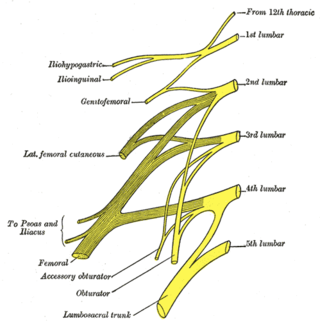
The lumbar nerves are the five pairs of spinal nerves emerging from the lumbar vertebrae. They are divided into posterior and anterior divisions.

<span class="mw-page-title-main">Ischium</span> Lower and back region of the hip bone

The ischium forms the lower and back region of the hip bone.

<span class="mw-page-title-main">Nerve to obturator internus</span> Human nerve

The nerve to obturator internus is a mixed nerve providing motor innervation to the obturator internus muscle and gemellus superior muscle, and sensory innervation to the hip joint. It is a branch of the sacral plexus. It is one of the group of deep gluteal nerves.

<span class="mw-page-title-main">Nerve to quadratus femoris</span>

The nerve to quadratus femoris is a nerve of the sacral plexus that provides motor innervation to the quadratus femoris muscle and gemellus inferior muscle, and an articular branch to the hip joint. The nerve leaves the pelvis through the greater sciatic foramen.

<span class="mw-page-title-main">Hip bone</span> Bone of the pelvis

The hip bone is a large flat bone, constricted in the center and expanded above and below. In some vertebrates it is composed of three parts: the ilium, ischium, and the pubis.

<span class="mw-page-title-main">Pelvis</span> Lower torso of the human body

The pelvis is the lower part of the trunk, between the abdomen and the thighs, together with its embedded skeleton.

<span class="mw-page-title-main">Gemelli muscles</span> Two small muscular fasciculi, accessories to the tendon of the internal obturator muscle

The gemelli muscles are the inferior gemellus muscle and the superior gemellus muscle, two small accessory fasciculi to the tendon of the internal obturator muscle. The gemelli muscles belong to the lateral rotator group of six muscles of the hip that rotate the femur in the hip joint.

References

  1. MedicalMnemonics.com: 833 3471 657
  2. 1 2 Bojsen-Møller, Finn; Simonsen, Erik B.; Tranum-Jensen, Jørgen (2001). Bevægeapparatets anatomi[Anatomy of the Locomotive Apparatus] (in Danish) (12th ed.). p. 365. ISBN   978-87-628-0307-7.