Ischial spine

Last updated
Ischial spine
Gray343.png
Capsule of hip-joint (distended). Posterior aspect. (Spine of ischium labeled at upper left.)
Gray341.png
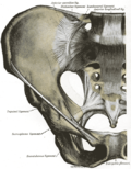
Left hip-joint, opened by removing the floor of the acetabulum from within the pelvis. (Spine of ischium labeled at center left.)
Details
Identifiers
Latin spina ischiadica
spina ischiaca
spina ischialis
TA98 A02.5.01.205
TA2 1343
FMA 17028
Anatomical terms of bone

The ischial spine is part of the posterior border of the body of the ischium bone of the pelvis. It is a thin and pointed triangular eminence, more or less elongated in different subjects.

Contents

Structure

PartAttachment
external surface gemellus superior muscle [1]
internal surface coccygeus muscle, [2] levator ani muscle, pelvic fascia
pointed extremity sacrospinous ligament

The pudendal nerve travels close to the ischial spine. [3]

Clinical significance

The ischial spine can serve as a landmark in pudendal anesthesia, as the pudendal nerve lies close to the ischial spine. [3] [4]

Additional images

Related Research Articles

<span class="mw-page-title-main">Pudendal nerve</span> Main nerve of the perineum

The pudendal nerve is the main nerve of the perineum. It is a mixed nerve and also conveys sympathetic autonomic fibers. It carries sensation from the external genitalia of both sexes and the skin around the anus and perineum, as well as the motor supply to various pelvic muscles, including the male or female external urethral sphincter and the external anal sphincter.

<span class="mw-page-title-main">Sartorius muscle</span> Longest muscle in the human body

The sartorius muscle is the longest muscle in the human body. It is a long, thin, superficial muscle that runs down the length of the thigh in the anterior compartment.

<span class="mw-page-title-main">Umbilical artery</span> Artery in the abdominal and pelvic regions

The umbilical artery is a paired artery that is found in the abdominal and pelvic regions. In the fetus, it extends into the umbilical cord.

<span class="mw-page-title-main">Spinal canal</span> Passage through the vertebral column containing the spinal cord

In human anatomy, the spinal canal, vertebral canal or spinal cavity is an elongated body cavity enclosed within the dorsal bony arches of the vertebral column, which contains the spinal cord, spinal roots and dorsal root ganglia. It is a process of the dorsal body cavity formed by alignment of the vertebral foramina. Under the vertebral arches, the spinal canal is also covered anteriorly by the posterior longitudinal ligament and posteriorly by the ligamentum flavum. The potential space between these ligaments and the dura mater covering the spinal cord is known as the epidural space. Spinal nerves exit the spinal canal via the intervertebral foramina under the corresponding vertebral pedicles.

<span class="mw-page-title-main">Quadriceps</span> Group of human leg muscle

The quadriceps femoris muscle is a large muscle group that includes the four prevailing muscles on the front of the thigh. It is the sole extensor muscle of the knee, forming a large fleshy mass which covers the front and sides of the femur. The name derives from Latin four-headed muscle of the femur.

<span class="mw-page-title-main">Coccygeus muscle</span> Muscle of the lower back arising by its apex from the spine of the ischium

The coccygeus muscle or ischiococcygeus is a muscle of the pelvic floor located posterior to levator ani and anterior to the sacrospinous ligament.

<span class="mw-page-title-main">Obturator internus muscle</span> One of six small hip muscles in the lateral rotator group

The internal obturator muscle or obturator internus muscle originates on the medial surface of the obturator membrane, the ischium near the membrane, and the rim of the pubis.

<span class="mw-page-title-main">Psoas minor muscle</span> Long, slender skeletal muscle located anterior to the psoas major muscle

The psoas minor muscle is a long, slender skeletal muscle. When present, it is located anterior to the psoas major muscle.

<span class="mw-page-title-main">Anterior superior iliac spine</span> Bony projection of the iliac bone

The anterior superior iliac spine (ASIS) is a bony projection of the iliac bone, and an important landmark of surface anatomy. It refers to the anterior extremity of the iliac crest of the pelvis. It provides attachment for the inguinal ligament, and the sartorius muscle. The tensor fasciae latae muscle attaches to the lateral aspect of the superior anterior iliac spine, and also about 5 cm away at the iliac tubercle.

<span class="mw-page-title-main">Femoral nerve</span> Long nerve down the thigh and inner leg

The femoral nerve is a nerve in the thigh that supplies skin on the upper thigh and inner leg, and the muscles that extend the knee. It is the largest branch of the lumbar plexus.

The gluteal tuberosity is the lateral one of the three upward prolongations of the linea aspera of the femur, extending to the base of the greater trochanter. It serves as the principal insertion site for the gluteus maximus muscle.

<span class="mw-page-title-main">Supraspinous ligament</span>

The supraspinous ligament, also known as the supraspinal ligament, is a ligament found along the vertebral column.

<span class="mw-page-title-main">Inferior gluteal artery</span>

The inferior gluteal artery is a terminal branch of the anterior trunk of the internal iliac artery. It exits the pelvis through the greater sciatic foramen. It is distributed chiefly to the buttock and the back of the thigh.

<span class="mw-page-title-main">Iliocostalis</span>

Iliocostalis muscle is the muscle immediately lateral to the longissimus that is the nearest to the furrow that separates the epaxial muscles from the hypaxial. It lies very deep to the fleshy portion of the serratus posterior muscle. It laterally flexes the vertebral column to the same side.

<span class="mw-page-title-main">Greater sciatic foramen</span> Part of the pelvis

The greater sciatic foramen is an opening in the posterior human pelvis. It is formed by the sacrotuberous and sacrospinous ligaments. The piriformis muscle passes through the foramen and occupies most of its volume. The greater sciatic foramen is wider in women than in men.

<span class="mw-page-title-main">Pelvic brim</span>

The pelvic brim is the edge of the pelvic inlet. It is an approximately Mickey Mouse head-shaped line passing through the prominence of the sacrum, the arcuate and pectineal lines, and the upper margin of the pubic symphysis.

<span class="mw-page-title-main">Iliac fossa</span>

The iliac fossa is a large, smooth, concave surface on the internal surface of the ilium.

<span class="mw-page-title-main">Nerve to obturator internus</span> Human nerve

The nerve to obturator internus is a mixed nerve providing motor innervation to the obturator internus muscle and gemellus superior muscle, and sensory innervation to the hip joint. It is a branch of the sacral plexus. It is one of the group of deep gluteal nerves.

<span class="mw-page-title-main">Esophageal plexus</span>

The esophageal plexus is formed by nerve fibers from two sources, branches of the vagus nerve, and visceral branches of the sympathetic trunk. The esophageal plexus and the cardiac plexus contain the same types of fibers and are both considered thoracic autonomic plexus.

<span class="mw-page-title-main">Facet joint</span> Joint between two adjacent vertebrae

The facet joints are a set of synovial, plane joints between the articular processes of two adjacent vertebrae. There are two facet joints in each spinal motion segment and each facet joint is innervated by the recurrent meningeal nerves.

References

PD-icon.svgThis article incorporates text in the public domain from page 235 of the 20th edition of Gray's Anatomy (1918)

  1. Chaitow, Leon; DeLany, Judith (2011-01-01), Chaitow, Leon; DeLany, Judith (eds.), "Chapter 12 - The hip", Clinical Application of Neuromuscular Techniques, Volume 2 (Second Edition), Oxford: Churchill Livingstone, pp. 391–445, doi:10.1016/b978-0-443-06815-7.00012-7, ISBN   978-0-443-06815-7 , retrieved 2021-02-19
  2. Bharucha, ADIL E.; Klingele, CHRISTOPHER J. (2005-01-01), Dyck, Peter J.; Thomas, P. K. (eds.), "Chapter 13 - Autonomic and Somatic Systems to the Anorectum and Pelvic Floor", Peripheral Neuropathy (Fourth Edition), Philadelphia: W.B. Saunders, pp. 279–298, doi:10.1016/b978-0-7216-9491-7.50016-8, ISBN   978-0-7216-9491-7 , retrieved 2021-02-19
  3. 1 2 Christo, Paul J.; Hobelmann, Greg (2009-01-01), Smith, HOWARD S. (ed.), "Chapter 29 - PELVIC PAIN", Current Therapy in Pain, Philadelphia: W.B. Saunders, pp. 216–227, doi:10.1016/b978-1-4160-4836-7.00029-8, ISBN   978-1-4160-4836-7 , retrieved 2021-02-19
  4. "Clinical Case - Perineum & External Genitalia". Archived from the original on 2011-07-17. Retrieved 2009-05-27.