Pelvic inlet

Last updated
Pelvic inlet
Pelvic inlet 03 animation.gif
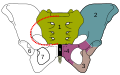
The pelvic inlet (shown in red)
Gray238.png
Diameters of superior aperture of lesser pelvis (female)
Details
Identifiers
Latin apertura pelvis superior
TA98 A02.5.02.008
TA2 1289
FMA 17272
Anatomical terms of bone

The pelvic inlet or superior aperture of the pelvis is a planar surface which defines the boundary between the pelvic cavity and the abdominal cavity (or, according to some authors, between two parts of the pelvic cavity, called lesser pelvis and greater pelvis). It is a major target of measurements of pelvimetry.

Contents

Its position and orientation relative to the skeleton of the pelvis is anatomically defined by its edge, the pelvic brim. The pelvic brim is an approximately apple-shaped line passing through the prominence of the sacrum, the arcuate and pectineal lines, and the upper margin of the pubic symphysis.

Occasionally, the terms pelvic inlet and pelvic brim are used interchangeably.

Boundaries

The edge of the pelvic inlet (pelvic brim) is formed as follows:

Anteriorly
by the pubic crest (or pubic symphysis)
Laterally
by the iliopectineal line
(same as other side)
Posteriorly
by the anterior margin of the base of the sacrum (or the ala of sacrum) and sacrovertebral angle (or sacral promontory)

Diameters

The diameters or conjugates of the pelvis are measured at the pelvic inlet and outlet and as oblique diameters.

NameDescriptionAverage measurement in female
Anteroposterior or conjugate diameter or conjugata veraExtends from the upper margin of the pubic symphysis to the sacrococcygeal joint;about 110 mm.
Transverse diameterExtends across the greatest width of the superior aperture, from the middle of the brim on one side to the same point on the opposite;about 135 mm.
Oblique diameterExtends from the iliopectineal eminence of one side to the sacroiliac articulation of the opposite side;about 125 mm.
Anatomical conjugateExtends from the pubic symphysis to the promontory;about 120 mm.
Diagonal conjugateExtends from lower margin of the pubic symphysis to the sacral promontory;about 130 mm.
Straight conjugateExtends from the lower border of the pubic symphysis to the tip of coccyx. The coccyx can bend posteriorly and expand the diameter with 25 mm;about 95 mm (+ 25 mm).
Median conjugateExtends from the lower border of the pubic symphysis to the lower border of the sacrum;about 115 mm.

Two diameters may be measured from the outside of the body using a pelvimeter

NameDescriptionAverage measurement in female
Interspinous distanceExtends between the anterior superior iliac spines;about 260 mm.
Intercristal distanceExtends between the furthest later points of the two iliac crests;about 290 mm.

Additional images

See also

Related Research Articles

<span class="mw-page-title-main">Peritoneum</span> Serous membrane that forms lining of abdominal cavity or coelom

The peritoneum is the serous membrane forming the lining of the abdominal cavity or coelom in amniotes and some invertebrates, such as annelids. It covers most of the intra-abdominal organs, and is composed of a layer of mesothelium supported by a thin layer of connective tissue. This peritoneal lining of the cavity supports many of the abdominal organs and serves as a conduit for their blood vessels, lymphatic vessels, and nerves.

<span class="mw-page-title-main">Abdominal cavity</span> Body cavity in the abdominal area

The abdominal cavity is a large body cavity in humans and many other animals that contains many organs. It is a part of the abdominopelvic cavity. It is located below the thoracic cavity, and above the pelvic cavity. Its dome-shaped roof is the thoracic diaphragm, a thin sheet of muscle under the lungs, and its floor is the pelvic inlet, opening into the pelvis.

<span class="mw-page-title-main">Sacrum</span> Triangular-shaped bone at the bottom of the spine

The sacrum, in human anatomy, is a large, triangular bone at the base of the spine that forms by the fusing of the sacral vertebrae (S1–S5) between ages 18 and 30.

<span class="mw-page-title-main">Sigmoid colon</span> Section of the large intestine closest to the rectum and anus

The sigmoid colon is the part of the large intestine that is closest to the rectum and anus. It forms a loop that averages about 35–40 centimetres (14–16 in) in length. The loop is typically shaped like a Greek letter sigma (ς) or Latin letter S. This part of the colon normally lies within the pelvis, but due to its freedom of movement it is liable to be displaced into the abdominal cavity.

<span class="mw-page-title-main">Pubic symphysis</span> Cartilaginous joint between the front of the left and right hip bones

The pubic symphysis is a secondary cartilaginous joint between the left and right superior rami of the pubis of the hip bones. It is in front of and below the urinary bladder. In males, the suspensory ligament of the penis attaches to the pubic symphysis. In females, the pubic symphysis is close to the clitoris. In most adults it can be moved roughly 2 mm and with 1 degree rotation. This increases for women at the time of childbirth.

<span class="mw-page-title-main">Pelvic floor</span> Anatomical structure

The pelvic floor or pelvic diaphragm is composed of muscle fibers of the levator ani, the coccygeus muscle, and associated connective tissue which span the area underneath the pelvis. The pelvic diaphragm is a muscular partition formed by the levatores ani and coccygei, with which may be included the parietal pelvic fascia on their upper and lower aspects. The pelvic floor separates the pelvic cavity above from the perineal region below. Both males and females have a pelvic floor. To accommodate the birth canal, a female's pelvic cavity is larger than a male's.

<span class="mw-page-title-main">Internal pudendal artery</span>

The internal pudendal artery is one of the three pudendal arteries. It branches off the internal iliac artery, and provides blood to the external genitalia.

<span class="mw-page-title-main">External iliac artery</span> Arteries of the pelvis

The external iliac arteries are two major arteries which bifurcate off the common iliac arteries anterior to the sacroiliac joint of the pelvis.

The obstetrical dilemma is a hypothesis to explain why humans often require assistance from other humans during childbirth to avoid complications, whereas most non-human primates give birth unassisted with relatively little difficulty. This occurs due to the tight fit of the fetal head to the maternal birth canal, which is additionally convoluted, meaning the head and therefore body of the infant must rotate during childbirth in order to fit, unlike in other, non-upright walking mammals. Consequently, there is a usually high incidence of cephalopelvic disproportion and obstructed labor in humans.

<span class="mw-page-title-main">Abdomen</span> Part of the body between the chest and pelvis

The abdomen is the part of the body between the thorax (chest) and pelvis, in humans and in other vertebrates. The abdomen is the front part of the abdominal segment of the torso. The area occupied by the abdomen is called the abdominal cavity. In arthropods it is the posterior tagma of the body; it follows the thorax or cephalothorax.

Pelvimetry is the measurement of the female pelvis. It can theoretically identify cephalo-pelvic disproportion, which is when the capacity of the pelvis is inadequate to allow the fetus to negotiate the birth canal. However, clinical evidence indicate that all pregnant women should be allowed a trial of labor regardless of pelvimetry results.

<span class="mw-page-title-main">Pubis (bone)</span> Most forward-facing of the three main regions making up the os coxa

In vertebrates, the pubic region is the most forward-facing of the three main regions making up the coxal bone. The left and right pubic regions are each made up of three sections, a superior ramus, inferior ramus, and a body.

<span class="mw-page-title-main">Pelvic cavity</span> Body cavity bounded by the bones of the pelvis

The pelvic cavity is a body cavity that is bounded by the bones of the pelvis. Its oblique roof is the pelvic inlet. Its lower boundary is the pelvic floor.

<span class="mw-page-title-main">Pelvic brim</span>

The pelvic brim is the edge of the pelvic inlet. It is an approximately Mickey Mouse head-shaped line passing through the prominence of the sacrum, the arcuate and pectineal lines, and the upper margin of the pubic symphysis.

<span class="mw-page-title-main">Linea terminalis</span>

The linea terminalis or innominate line consists of the pubic crest, pectineal line, the arcuate line, the sacral ala, and the sacral promontory.

<span class="mw-page-title-main">Pelvic fascia</span>

The pelvic fasciae are the fascia of the pelvis and can be divided into:

<span class="mw-page-title-main">Pelvic outlet</span>

The lower circumference of the lesser pelvis is very irregular; the space enclosed by it is named the inferior aperture or pelvic outlet. It is an important component of pelvimetry.

<span class="mw-page-title-main">Hip bone</span> Bone of the pelvis

The hip bone is a large flat bone, constricted in the center and expanded above and below. In some vertebrates it is composed of three parts: the ilium, ischium, and the pubis.

<span class="mw-page-title-main">Pelvis</span> Lower part of the trunk of the human body between the abdomen and the thighs

The pelvis is the lower part of the trunk, between the abdomen and the thighs, together with its embedded skeleton.

X-rays of hip dysplasia are one of the two main methods of medical imaging to diagnose hip dysplasia, the other one being medical ultrasonography. Ultrasound imaging yields better results defining the anatomy until the cartilage is ossified. When the infant is around 3 months old a clear roentgenographic image can be achieved. Unfortunately the time the joint gives a good x-ray image is also the point at which nonsurgical treatment methods cease to give good results.